StudentShare
Contact Us
Sign In / Sign Up for FREE
Search
Go to advanced search...
Free

Dermatological Conditions: Acne Ulgaris, Rosacea, Eczema, and Seborrheic Dermatitis - Assignment Example

Cite this document
Summary
"Dermatological Conditions: Acne Ulgaris, Rosacea, Eczema, and Seborrheic Dermatitis" paper examines dermatological conditions associated with the skin which forms the largest portion of the human body. Essentially, it is pertinent to categorize and classify common skin lacerations…
Download full paper File format: .doc, available for editing
GRAB THE BEST PAPER91.5% of users find it useful
Dermatological Conditions: Acne Ulgaris, Rosacea, Eczema, and Seborrheic Dermatitis
Read Text Preview

Extract of sample "Dermatological Conditions: Acne Ulgaris, Rosacea, Eczema, and Seborrheic Dermatitis"

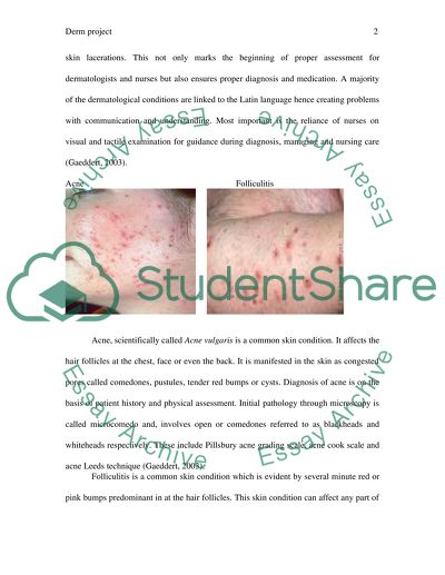
? Derm project Derm Project Dermatological conditions are associated with the skin which forms the largest portion of the human body. Essentially, it is pertinent to categorize and classify common skin lacerations. This not only marks the beginning of proper assessment for dermatologists and nurses but also ensures proper diagnosis and medication. A majority of the dermatological conditions are linked to the Latin language hence creating problems with communication and understanding. Most important is the reliance of nurses on visual and tactile examination for guidance during diagnosis, managing and nursing care (Gaeddert, 2003). Acne Folliculitis Acne, scientifically called Acne vulgaris is a common skin condition. It affects the hair follicles at the chest, face or even the back. It is manifested in the skin as congested pores called comedones, pustules, tender red bumps or cysts. Diagnosis of acne is on the basis of patient history and physical assessment. Initial pathology through microscopy is called microcomedo and, involves open or comedones referred to as blackheads and whiteheads respectively. These include Pillsbury acne grading scale, acne cook scale and acne Leeds technique (Gaeddert, 2003). Folliculitis is a common skin condition which is evident by several minute red or pink bumps predominant in at the hair follicles. This skin condition can affect any part of the body including the back, arms, legs, chest and cheeks. Ranging between 10 - 100 bumps, this skin condition appears as a scatter within the affected area. The spots or bumps can be slightly red indicating an inflammation. Accidental scratching of the bump reveals a coiled hair engulfed within the bump. Diagnosis of Folliculitis basically involves the assessment of the skin’s appearance (Gaeddert, 2003). On rare instances is skin biopsies required so as to help the dermatologist on diagnosis. Additionally, the skin could be cultured in bacterial media so as to help in detection of the causative agent. Microscopic tests on the skin including fungal tests can be conducted using potassium hydroxide. This procedure helps in determining whether the condition is caused by a fungus or yeast. A histopathological procedure can be conducted to reveal epidermal hyperkeratosis as well as clustering of leukocytes. Rosacea Eczema Rosacea is a thriving skin condition which affects the eyes and the face. It is characterized by reddening of the affected region indicating a possible inflammation as well as emergence of pimples. This dermatological condition is most pronounced in women and individuals with fair skin tone. Usually, Rosacea is evident in the age range of between 30 to 60 years. Also termed as acne rosacea, this condition is characterized by symptoms of swollen nose, thick skin, red and itchy eyes. Moreover, there is a stinging facial skin on application of lotions as well as minute red veins on the face. The diagnosis of Rosacea is mostly dependent on physical examination categorized as primary or secondary features. A patient is diagnosed with Rosacea if he or she possesses one of the primary features of persistent flushing, pimples, visible blood vessels. Secondary features are manifest in several subtypes including Papulopustular, Phymatous Erythematotelangiectatic and ocular rosacea (Gaeddert, 2003). Eczema is characterized by vesicles that are small and which contain fluid substance. These vessicles are pimple like and are evident as reddish and swollen. When dried, the fluids leave behind dry patches on the surface of the affected areas. This condition occurs in the facial region in a majority of patients. Eczema can be diagnosed through microscopy. Seborrheic Dermatitis Scalp psoriasis A close relative psoriasis is the seborrheic dermatitis that affects the scalp as well. This condition is inflammatory and it leads to the production of flaky, white to yellowish scabs on the skin. Symptoms of this condition include reddish swollen patches around the nose, armpits and mouth. Others include itching, widespread scabs and a flaky skin which is oily and waxy. Seborrheic dermatitis can be diagnosed through visual check and a medical history analysis. Skin biopsies can also be done to eliminate the probability of other related conditions (Gaeddert, 2003). Scalp psoriasis is a noncommunicable disease with a genetic predominance. This disease emanates from the immune system and affects mostly the skin and the joints. This condition can be mild although accompanied with minor superior scaling. Scalp psoriasis can be very acute as characterized with thick layered plaques covering the whole scalp. Additionally, psoriasis can elongate beyond the hairline onto the forehead, neck region and around the ears. There is no single diagnostic approach for detection of psoriasis and, it is therefore important that the doctor distinguishes this condition from seborrheic dermatitis (Gaeddert, 2003). Pityriasis Rosea Erythema Multiforme Pityriasis Rosea is a skin condition that is manifested by the appearance of scatter redness. It is linked to a very slight scaling of the skin and, is one of the placid form of dermatoses. This condition is also characterized by the upsurge of a single outsized plate and several minute plates on the skin portion affected. It is manifested by placid and intermittent itching, sore throat and nausea. Pityriasis rosea is diagnosed through visual examination of the skin. In this, there is usually a herald patch which is symmetrical and assumes a christmas tree patch afterwards. Alternatively, an elimination procedure involving potassium hydroxide can be performed. This is called the quick prep fungal test for detecting fungal infections. Additionally, blood test called rapid plasma reagent test can be performed to identify secondary syphilis. In rare occasions, skin biopsies are performed to distinguish fungus and other irritations (Gaeddert, 2003). Erythema is a dermatological condition that is manifested with reddening of the skin. Known an Erythema Multiforme, this condition is a hypersensitivity response that is triggered as a result of infections predominantly herpes simplex virus. Erythema occurs as a typical target lesion and, may incorporate mucous membrane (Gaeddert, 2003). Herpes Simplex virus Phytophotodermatitis  The main causative agent of HSV is the HSV-1 which occurs mostly around the lips and mouth. HSV-2 is the main agent of causing genital herpes infection. This variant of herpes is transmitted mainly through sexual intercourse. Herpes simplex virus 1 is manifested with painful blisters filled with liquid on the affected region. Other symptoms are red swollen blisters around the thighs and swollen lymph nodes. Herpes condition is diagnosed by visual assessment of the blisters. In some instances, blood tests are conducted to ascertain the actual infection. Phytophotodermatitis is a skin condition that majorly affects the cutaneous layer of this body organ. It results from phototoxic inflammatory upsurge from long wave UV rays. It is characterized by blisters or lesions and it affects the regions exposed to the radiation. Diagnosis is dependent on linear streaks of hyperpigmentation as well as the inflammatory patterns. Seborrheic keratosis Dermatosis papulosa nigra Seborrheic keratosis is another skin condition that is characterized by a growth or a tumor-like cell. It is a non-cancerus growth that can increase in size to become thick and warty. The condition is common among the middle-aged and mature persons and, is not contagious at all. In a majority of the cases, the condition is depicted by a waxy look resembling the blob of warm. Seborrheic keratosis can appear in a any region of the skin. Seborrheic keratosis can be diagnosed through physical examination. Other cases may involve the removal of the growth for microscopic analysis (Gaeddert, 2003). Dermatosis papulosa nigra resemble seborrheic keratosis and is also a cutaneous benign conditioncommon among the blacks. it is characterized by small, multiple papules which are hyperpigmented on the face. It can be diagnosed through physical examination of the papules. Keratoacanthoma Cutaneous Horn Keratoacanthoma is a skin condition which has a relatively lower scale of malignancy emanating from the piolosebaceous glands. This skin condition is almost similar to squamous cell carcinoma. It is characterized by a drastic growth within the first few weeks. It is believed to be caused by carcinogenic substances as well as the sunlight. Keratoacanthoma grows between 1 to 2 centimetres in the first weeks before evolving into a lasting scar. The lesions occur in singlets and are firm in terms of texture. They are round in shape and appear as reddish papules which develop a dome shape. Keratoacanthoma is diagnosed via clinical and patient history procedure. Skin biopsies are also important in the diagnosis of this skin condition. Cutaneous horn is characterized by a conical elongationon the skin’s surface resembling a minute horn. It is distibuted on the pinna, face, nose or forearms. It is a hyperkeratotic papule measuring upto one centimetre in height from the basal side. Its diagnosis is basically based on visual assessment because it is prominent and distinct. Actinic keratosis Porokeratosis Actinic keratosis is a dermatose that develops upon long term exposure to sunlight. it is characterized by a rough, patch filled with scales. It affects the lips, ears, face, arms, neck and hands. Referred to as also as solar keratosis, this condition has a symptom of a small patch on the skin. It may be flat or raised on the skin surface and it,s color ranges from red brown to pink. Diagnosis of actinic keratosis is conducted thoroughly and involves a biopsy. Porokeratosis is a disoder that arises from clones during keratinization. It is manifested by a single or multiple patches surrounded by hyperkaratotic layer called cornoid lamella. It is characterized by reddish browm scabs and keratotic papules. It is diagnosed by dermascopy to reveal a white track structure with a brownish hue. (Gaeddert, 2003). Squamous Cell Carcinoma Warty dyskeratoma Squamous cell a carcinoma is a variant cancer of the skin that is caused by non-melanocytes. It is the second most common skin cancer after basal cell carcinoma.squamous cell carcinoma accounts for almost 20% of malignant cell in the cutaneous region. It occurs on the ears, neck, hands and the face. The major sigh of this non-melanocyte cancer is a developing bump which may have a rough surface filled with scabs and reddish patches. Any sore within the stated regions that does not heal could be a sign of squamous cell carcinoma. It is diagnosed through visual examination putting into consideration the texture, size, shape and color of the patch. Skin biopsies are also viable in the diagnosis of the condition. Warty dyskeratoma is an epidermal outgrowth of benign cells manifested as an umblicated lesion. It occurs mainly on the neck, face or head and is characterized by reddish lesions appearing as keratotic nodule. It is diagnosed via immunohistochemical techniques such as SERCA2 staining. SCC –in situ Merkel Cell Carcinoma  The other variant of Squamous cell carcinoma occurs in its original place hence termed in situ. This is because, the cell have not invaded or moved into a new location in the neighboring tissues or organs. Squamous cell carcinoma in situ is also called the Bowen’s disease and is progressive and persistent in nature. It has the traits of a raised reddish broken patch and is evident within the mucosal regions. It can be diagnosed via microscopic analysis for the arrangement of the cells (Gaeddert, 2003). Merkel cell is presented as a solitaryand firm nodule. This lesion is always more than 2 centimetres but can surpass 15 centimetres in size. The lesions are normally purple in color and are depicted as shiny plaques. It is diagnosed through microscopy of the affected tissues (Gaeddert, 2003). Basal Cell Carcinoma Syringoma Basal cell carcinoma is the leading non-melanocyte skin cancer in the entire world. Basal cell carcinomas occur as tumors when there is an exposure to sunlight. These tumors develop at a lower rate compared to those of Squamous cell carcinoma. The metastasis rate of these cells is less than 1% and, mostly is predominant in the head region. The tumors tend to have a flat shape with a hard texture. The tumors are small and are slightly elevated on the skin surface. Additionally, they are shiny and translucent. In cases of minor accidents, these areas are prone to bleeding. The size of the tumors can range from a few millimeters to centimeters with regard to the diameter. In its diagnosis, skin biopsy remains relevant for confirmation and determination of subtypes. Equally important in the diagnosis is a punch biopsy when obtaining the thick sample after shave biopsy. Syringoma is characterized by multiple, minute yellow papules in the lower eyelids or upper portion of the cheeks. The lesions appear as translucent, round or flat and are usually small in size. Syringoma is diagnosed via physical examination to differentiate it from other variants of carcinomas. Malignant melanoma Lentigo Malignant melanoma is a skin cancer variant that develops from the melanocytes hence it is a melanocytic cancer. These conditions are categorized in different classes from A to E. Characterized by scaly, itchy moles, malignant, melanoma remains a concern to humanity. Cancerous melanomas occur essentially in every part of the body but majorly within the skin surfaces. Several forms of malignant melanoma have been classified and include nodular, lentigo and superficial spreading melanomas. Malignant melanomas are diagnosed by physical examination of the spot and by carrying out a biopsy. Another technique clinical technique called epiluminescence microscopy can also be conducted during diagnosis (Gaeddert, 2003). Lentigo is a skin condition that is small and distinctively enclosed as a macule. It has a dark pigmentation and it assumes an oval or round shape measuring between 3- 14 milimetres. Lentigo is diagnosed through dermatoscopy and excision of the affected region for biopsy (Gaeddert, 2003). Reference Gaeddert, A. (2003). Healing Skin Disorders: Natural Treatments for Dermatological Conditions. California: North Atlantic Books,. Read More
Cite this document
  • APA
  • MLA
  • CHICAGO
(“Derm Project Assignment Example | Topics and Well Written Essays - 1000 words”, n.d.)
Derm Project Assignment Example | Topics and Well Written Essays - 1000 words. Retrieved from https://studentshare.org/nursing/1439481-derm-project
(Derm Project Assignment Example | Topics and Well Written Essays - 1000 Words)
Derm Project Assignment Example | Topics and Well Written Essays - 1000 Words. https://studentshare.org/nursing/1439481-derm-project.
“Derm Project Assignment Example | Topics and Well Written Essays - 1000 Words”, n.d. https://studentshare.org/nursing/1439481-derm-project.
  • Cited: 0 times

CHECK THESE SAMPLES OF Dermatological Conditions: Acne Ulgaris, Rosacea, Eczema, and Seborrheic Dermatitis

Atopic Eczema or Atopic Dermatitis: a Chronic Relapsing Inflammatory Skin Condition

This essay "Atopic Eczema or Atopic dermatitis: a Chronic Relapsing Inflammatory Skin Condition" focuses on the research studies conducted to find out the efficiency of prevailing treatments.... Among the children affected, about 65% developed the symptoms of Atopic eczema within their first year of life and 85% were affected with it before they were 5 years old (Atopic dermatitis, 2011).... Corticosteroid creams and ointments and immunosuppressive drugs are the chosen treatments for atopic eczema....
14 Pages (3500 words) Essay

Pathophysiology of Eczema

The paper "Pathophysiology of Eczema" pinpoints the exact cause of eczema and dermatitis is not clearly known, but it may be a result of interplay of host, genetic and environmental factors.... Eczema or dermatitis is a disorder in which the skin is inflamed (especially epidermis) along with the development of vesiculation sometimes in acute phases.... Some of the common types include:-• Allergic contact eczema – Following contact with a foreign substance such as a poison, lotion, etc, there is a local allergic reaction• Atopic dermatitis – It is a chronic inflammatory skin disorder and often accompanied by other disorders such as asthma and hay fever....
6 Pages (1500 words) PowerPoint Presentation

Atopic Dermatitis: Eczema

(Bieber & Leung, 2002)Differences between eczema and atopic dermatitis There are different categories of eczema such as contact, irritant, and allergic eczema which may be similar to atopic dermatitis or not.... This research paper "Atopic dermatitis: Eczema" explores atopic dermatitis as a non-contagious skin disease it causes itchy skin.... The skin in atopic dermatitis becomes inflamed and itchy, it becomes red and swells up and may cause small blisters, crusting, scaling, cracking, or weeping of the skin....
6 Pages (1500 words) Research Paper

Eczema: Diagnosis and Treatment

seborrheic dermatitis (aka cradle cap in infants, dandruff) causes dry or greasy scaling of the scalp and eyebrows.... atopic dermatitis) is thought to be hereditary, and often runs in families whose members also have hay fever and asthma.... Contact dermatitis is of two types: allergic (resulting from a delayed reaction to some allergen, such as poison ivy, or nickel), and irritant (resulting from direct reaction to, say, a solvent).... dermatitis herperiformis causes an intensely itchy and typically symmetrical rash on arms, thighs, knees, and back....
5 Pages (1250 words) Essay

Clinical Dermatological Conditions: Psoriasis

This essay "Clinical dermatological conditions: Psoriasis" is about one of the most dermatological conditions common and recent evidence points to an etiology encompassing systemic, immunological, autoimmune, and genetic elements, but the exact mechanism for its initiation is not known.... According to Nickoloff and Nestle (2004), approximately 30% of patients suffering from psoriasis attempt suicide, which places the disease on par with other major medical conditions such as depression, heart disease, and diabetes in terms of psychological impact....
22 Pages (5500 words) Essay

Impact of Dermatology Conditions

acne vulgaris is a common chronic inflammatory dermatological condition that affects more than 90% adolescents during their teenage growth spurts due to the surge in hormones and often continues well into the adulthood.... acne (also called as pimples, zits, blemishes) mostly tend to appear on face, neck, upper chest, shoulders and back.... Socially, acne has been a source of low self esteem, shame, embarrassment and often depression.... Even though the pattern of inheritance has not been identified yet, but 3 of 4 children are likely to have acne if both parents happen to suffer from the disease....
4 Pages (1000 words) Essay

Causes, Treatment and Prevention of Eczema

The range of severity of disease is broad, the causes are multiple and complex, and treatment approaches involved immunosuppressant and anti-inflammatory pharmacologic agents must be tailored to the type of eczema and its clinical severity in each individual patient.... When it is caused by contact dermatitis, the exposed area is the site where eczema develops.... The paper will review the most common types of eczema, their causes and the types of treatments that are most useful in the management of this disease....
6 Pages (1500 words) Research Paper

Effectiveness of Using Low-Intensity Laser Therapy

rosacea mostly affects the skin around the face and in very rare instances does the skin on the other parts of the body affected.... The constant flushing is caused by a condition called vascular rosacea which is characterized by enlargement of blood vessels under the facial skin which can sometimes be visible as small red lines.... nother condition of rosacea is called inflammatory rosacea and is characterized by constant redness of the skin swellings containing pus....
7 Pages (1750 words) Essay
sponsored ads
We use cookies to create the best experience for you. Keep on browsing if you are OK with that, or find out how to manage cookies.
Contact Us