StudentShare
Contact Us
Sign In / Sign Up for FREE
Search
Go to advanced search...
Free

Diffusion MRI Stimulation theory - Admission/Application Essay Example

Cite this document
Summary
The paper “Diffusion MRI Stimulation theory” will analyze the scenario in which the walk of molecules abides by the principles of the Brownian particle’s elements. The fundamental example essentially describes the phenomenon by depicting the occurrence of the process…
Download full paper File format: .doc, available for editing
GRAB THE BEST PAPER95.2% of users find it useful
Diffusion MRI Stimulation theory
Read Text Preview

Extract of sample "Diffusion MRI Stimulation theory"

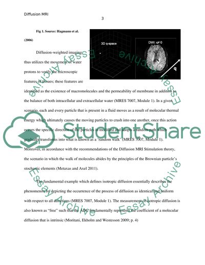
Diffusion MRI Stimulation theory The occurrence of self-diffusion of water molecules was first observed by Robert Brown in 1827, this process is characterized by continuous random thermal motion which all molecules above the temperature of zero degrees kelvin experience (Johansen-Berg and Behrens 2009; p. 38). Another definition of this phenomenon has been described by Luna, Ribes and Soto (2012; p. 1) who state that molecules having a temperature of 0°K (-273°C) undergo a random notion which is known as the Brownian movement. Furthermore, the rate at which the molecules move during the occurrence of diffusion is essentially dependent upon kinetic energy which in turn relates to the temperature of the molecule (Luna, Ribes and Soto 2012; p. 1). According to Koh and Thoeny (2010; p. 19) diffusion-weighted imaging has been one of the most pivotal advancements with regards to the progress of neurological imaging procedures in the past 10 years. This method is employs a chronological sequence to establish the microscopic random movement of water protons (Bammer 2003) and has thus been employed in the examination of numerous diseases due the various benefits that can be obtained as a consequence of its initiation (Kono et al. 2001). Perhaps, the most valued advantage of diffusion-weighted imaging is that enables technicians to save considerable amount of time by providing a single image which is required for the purpose (Hagmann et al. 2006). The nature of the procedure permits its implementation in a host of routine procedures, for example, in the case of acute stroke the enlargement of local cell causes the hindrance of water movement to enhance as a ramification of which a vivid display may appear in the location of lesion (Hagmann et al. 2006). Diffusion-weighted imaging thus utilizes the movement of water protons to verify the microscopic features of tissues; these features are identified as the existence of macromolecules and the permeability of membrane in addition to the balance of both intracellular and extracellular water (MRES 7007, Module 1). In a given scenario, each and every particle that is present in a fluid moves as a result of molecular thermal energy which ultimately causes the moving particles to crash into one another, once this action occurs the specific direction of the particles in modified and adopts a random path which gradually transforms into what is known as a ‘random walk’ (MRES 7007, Module 1). Moreover, in accordance with the recommendations of the Diffusion MRI Stimulation theory, the scenario in which the walk of molecules abides by the principles of the Brownian particle’s stochastic elements (Metaxas and Axel 2011). The fundamental example which defines isotropic diffusion essentially describes the phenomenon by depicting the occurrence of the process of diffusion as identical and uniform with respect to all directions (MRES 7007, Module 1). The measurement of isotropic diffusion is also known as “free” such that the ADC fundamentally represents the coefficient of a molecular diffusion that is intrinsic (Moritani, Ekholm and Westesson 2009; p. 4) . As per this case, the measurement of diffusion is conducted on the basis of the following formula: In the aforementioned formula, N reflects the dimension of molecular movement whereas; D is prescribed as the coefficient of diffusion at a certain temperature which is represented by t. Moreover, r2 defines the distance that has been covered by the molecule as a result of its movement. Peled et al. (1999) acknowledge the issue of restricted diffusion which may emerge as a result of the presence of barriers in tissues that are responsible for creating a correlation between a diffusion coefficient, the time it takes for diffusion to occur and in some cases the strength of the gradient. The scope of this observation is indicative of the presence of three barriers which are categorized as; permeable, non-permeable and partially permeable (MRES 7007, Module 1; Alarcon and Valentin 2012; p. 259). A permeable boundary allows the occurrence of free diffusion, on the contrary, in a non-permeable case a molecule is reflected once it meets with the barrier (MRES 7007, Module 1). Johansen-Berg and Behrens (2009; p. 132) claim that in case of membranes, which are only partially permeable, the impact of their displacement is enhanced over a giver period of time. Fig 2. This diagram depicts the Stejskal-Tanner sequence which is executed for the purpose of receiving DW images. Source: King (2006) When the first gradient of the sequence is executed, the position of stationary spins is determined or regulated in accordance to their position; consequently, this phase shift also shares a correlation with the gradient. During the process of 180 refocusing pulse the prior gradient is reversed and the same spin in the next scenario offsets the previous result to arrive at zero. It should be noted that the diffusion gradient does not only impact the stationary spins, it can also garner the same effect on the moving spins. This observation implies that when the first gradient is applied to the stationary spin, the phase shifts will be determined in accordance with their location, which in turn would be associated with the gradient’s amplitude. In the case of a refocusing pulse which is 180 the associated phase shift is essentially repealed (MRES 7007, Module 1). Consequently, when the diffusion gradient is applied to the second lobe the spins would generate an outcome that would counterbalance the phase, as a result of which the result that is achieved would be zero. However, in the scenario of moving spins the diffusion gradient’s first lobe would be responsible for creating phase shifts and the resultant scenario would cause the phase shifts to be reversed given the refocusing pulse of 180. Accordingly, in the case of the second lobe the resultant phase shifts would be dissimilar to that of the first lobe a ramification of which is characterized by the diminishment of the signal (MRES 7007, Module 1). Fig 2.1 This diagram shows the shift in phase for stationary spin and moving spins. The image shows the zero net result and the diminishment in signal for stationary spin and moving spin, respectively. (MRES 7007, Module 1). Source: Magnetic Resonance Imaging course at university of Queensland, (mres7007, module 1). The Skejskal-Tanner sequence displays the method in a series of sequences as presented in the diagram below: Fig 3. Spin echo sequence showing four distinct diffusion weighting schemes which are identified as a) constant gradient b) bipolar gradient c) several bipolar gradients d) Skejskal-Tanner sequence (Source: MRES 7007, Module 2). According to Johansen-Berg and Behrens (2009; p. 131), the incidence of hindered diffusion in brain tissue shares a relationship with the diffusion time and the mean displacement that is squared. This observation fundamentally implies that the distance which has been covered by a molecule is governed by two factors which are; the membrane’s degree of permeability and the diffusion coefficient which is referred to as the apparent diffusion coefficient (ADC) (MRES 7007, Module 1). King (2006; p. 103) claims that the Stejskal-Tanner sequence which is also known as the pulsed-gradient spin echo scheme is a widely adopted diffusion sequence for the purpose of achieving DWIs. The implementation of the Stejskal-Tanner sequence follows the utilization of two gradients which are located and arranged at a 180 degree pulse for refocusing to obtain a diffusion weighting that is controlled (King 2006; p. 103). According to King (2006; p. 103) the equation for determining the b-value is as follows: An analysis of this equation reveals that the signal intensity for MR is directly correlated to the b-value itself, this notion stipulates that an increase or decrease on the signal intensity of MR would have the same affect on the b-value, similarly, another observation with regards to the b-value postulates that a greater b-value would coincide with an enhancement in the quality of approximate diffusion tensor (King 2006; p. 103). Types of Diffusion: The process of diffusion can be categorized under isotropic diffusion and anisotropic diffusion. Moreover, isotropic diffusion can also be subcategorized under linear isotropic diffusion and non-linear isotropic diffusion. In linear isotropic diffusion, the diffusion function is marked by the constant presence of D; however, in non-linear isotropic diffusion the D shares a relationship with the local structure of an image (Torres and Sanfeliu 2000; p. 73). However, in basic terminology and as stated previously, isotropic diffusion represents a phenomenon in which the process of diffusion occurs uniformly and identically across all directions which essentially is indicative of the fact that the impact of a signal and the execution of a diffusion gradient would not produce varying results (Miller and Cummings 2007; p. 180; MRES 7007, Module 2). On the contrary, anisotropic diffusion presents a situation in which the diffusion of molecules occurs more in one direction in comparison with other directions; furthermore, this phenomenon is also marked by the presence of a flow (Miller and Cummings 2007; p. 180). The impact of isotropic diffusion and anisotropic on the MR signal is varied as a consequence of which the measurements for each type of diffusion are also different. McRobbie et al. (2006) note that the measurement of anisotropic diffusion is performed by the implementation of diffusion weighting that must occur in a minimum of 3 varying axes. The diagram below depicts the impact of this observation on a brain. Fig 4. This figure shows the DW images which have been acquired for a normal functioning brain for 3 varying axes, furthermore, the picture also presents a map of ADC in addition to its trace. Source: McRobbie et al. (2006) The phenomenon of isotropic diffusion performs in a uniform and identical manner in all directions while, in anisotropic diffusion the movement of molecules is more attracted towards a particular direction in comparison with others. Due to these features, the width of an isotropic medium remains the same or shows and delivers consistency in all directions which is depicted by the formation of a sphere. On the other hand, in anisotropic diffusion the lack of uniformity with regards to the direction leads to the creation of an ellipsoid (MRES 7007, Module 1). According to Toga and Mazziotta (2002; p. 383), in the case of anisotropic diffusion the measurements of MR are reliant upon the axis which has been utilized. Fig 5. Isotropic and Anisotropic Diffusion Representations Source: MRES 7007, Module 1 It is important to understand that the application of a Stjeskal and Tanner sequence, though widely popular and common in the attainment of diffusion-weighted images is not the only method which can be employed to achieve this aim. This notion postulates that other sequences can also be explored and adopted to obtain desired results. 1. Constant Diffusion Gradient According to Gillard, Waldman and Barker (2005; p. 56), this sequence requires the implementation of a spin echo and in this case the phase which has been acquired before the 180 RF will coincide with the RF which is acquired after due to the inverting principle. This notion points towards the occurrence of a zero phase offset. Moreover, the b-value is obtained through the following method (MRES 7007, Module 3): Fig 6. A Spin Echo Sequence in a Constant Diffusion Gradient Source: Gillard, Waldman and Barker (2005) 2. Bipolar Diffusion Gradients Unlike a Constant Diffusion Gradient, this method allows the implementation of both spin echo sequence and a gradient echo sequence. This technique may make use of two gradients, which are known as a positive gradient and a negative gradient to launch a diffusion weighting (Mori and Tournier 2013). It is important to state that this approach is also characterized by the presence of limitation which may limit the advantages that are gained as a consequence of its application. Hence, the two fundamental drawbacks of this technique are; the possibility of the loss of signal strength and deterioration of pulses which are shared by a pair of gradients (Mori and Tournier 2013). In the aforementioned technique, the b factor governs the diminishment of the signal which is as follows (MRES 7007, Module 3). Fig 6.1 As per the diagram, zero phase shifts are experienced by stationary spins in comparison with the scenario in which the bipolar gradient is implemented, on the other hand, moving spins are not characterized by zero phase shifts. Source: Magnetic Resonance Imaging course at university of Queensland, (MRES7007, Module 1) In the case of a SE sequence, it is plausible to implement a pair of bipolar gradients or numerous bipolar gradients. The latter has the ability to significantly diminish the time it takes for diffusion to occur. Both scenarios however, pose various limitations that must be considered before either option is selected. For example, the use of two bipolar gradients can be time-consuming on the other hand the use of numerous bipolar gradients adversely impacts precision (MRES 7007, Module 3). As suggested by the formula below, the implementation of several bipolar gradients is not responsible for minimizing the time it takes for diffusion to occur: Fig 6.2 Spin Echo Sequence Depicted with Bipolar Diffusion Gradients Source: Magnetic Resonance Imaging course at university of Queensland, (MRES7007, Module 1, 2 & 3). Fig 6.3 Enhancing the value of b without causing a reduction in diffusion time by the usage of several bipolar gradients Source: Magnetic Resonance Imaging course at university of Queensland, (MRES7007, Module 1, 2 & 3). 3. Diffusion Weighted Stimulated Echo This approach is implemented which require long diffusion in order to assess and examine slower rates of diffusion. As a result this method permits longer TE as well because the RF pulse accumulates magnetization (MRES 7007, Module 3). 4. Steady State Free Procession Sequences This approach functions by the utilization of RF pulses which are of a low flip angle as a result of which several echoes are developed. In this approach the gradient strength remains normal in comparison with other sequences which is why the rate of diffusion is also higher in comparison with other techniques. However, it should be noted that there are certain measures which can be taken to reduce the signal strength. One of the benefits of adopting this sequence is that it may result in the generation of high-quality images as suggested by the conclusions of various researches (Reiser, Semmler and Hricak 2008; p. 1245). Motion Artifacts According to Reiser, Semmler and Hricak (2008; p. 140) diffusion-weighted images maybe subjected to the emergence of motion artifacts due to the presence of various irregularities in the stage of phase formation, specifically when the raw data that has been obtained is of a multidimensional nature. Additionally, the source of these artifacts can also be traced to obtaining more numerous shots in a given time period (Johansen-Berg and Behrens 2009; p. 23). Other factors which may cause distortion of images include eddy currents (MRES 7007, Module 3). As the phenomenon of motion artifacts is essentially linked to signal sensitivity to large motions as suggested by Brown and Semelka (2011; p. 163), the b-value governs the nature of this relationship. The sequence which is most likely to be subjected to this phenomenon is the conventional diffusion weighted sequence, in order to reduce the impact of motion artifact it is recommended that certain measures be adopted, these techniques comprise of gating and navigator echoes amongst others (MRES 7007, Module 3). As noted by Brown and Smelka (2011; p. 140) the adoption of a navigator echo allows the measurement of an MR signal which is directed from numerous tissues, once measured the navigator echo has the capability to detect that motion has taken place. Another method that could be executed for tackling with this issue is that of single shot echo planar imaging (EPI) which is characterized by a steady and constant shift of image (Chen 2009; p. 22). The benefit of this technique is highlighted in its speed which has the capability to advance the accuracy and quality of image that is obtained. Measurement of Diffusion Information: The reasons that emphasis upon the significance of diffusion information essentially state that even when images have been acquired, the full extent of information cannot be extracted unless other procedures are initiated. Measurement of diffusion information can be conducted in for both the types of diffusion, that is isotropic diffusion and anisotropic diffusion. In case of isotropic diffusion, the direction or movement progresses to a single uniform point which is why the ADC in this scenario remains the same. On the other hand, for anisotropic diffusion the number of axes required amounts to at least three as in this system one particular direction is favored over others as a consequence of which the information obtained through ADC in this regard is limited (MRES 7007, Module 3). Diffusion Tensor: As stated above the extent of information that is gained in the case of anisotropic diffusion is limited and requires further development. To solve the issues that are associated with its limitation, a diffusion tensor can be employed. The diagram presented below depicts the matrix of this approach. Fig 7. A diffusion tensor matrix Source: MRES 7007, Module 3 In this 3x3 matrix, the process of diffusion is described by the employment of three distinct facets. In this case, Dxx, Dyy and Dzz are values which portray diffusion mobility. If the measurement of D is to presented, then this action would require the presence of at least six particular measurements. To achieve this aim, the diffusion gradients need to be obtained through orthogonal directions (MRES 7007, Module 3). Henceforth, the measurement of ADC can be summarized through the representation of various steps that must be fulfilled in order to achieve desired results. The first step requires the setting of standards and making appropriate amendments by modifying gradient coil, secondly, an image that has to obtained however it must be ensured that it does not have a diffusion gradient. Moreover, continuous scans could be acquired by keeping constant b-value with varying directions and vice versa. Once this action has been taken any distortions such as motion artifacts must be resolved by taking pertinent measures that have been presented previously. While, the aforementioned steps are indeed pivotal the most significant step relates to the calculation of the b-value which includes the presence of diffusion gradients. The last step of the process involves an examination of the diffusion tenor. Another important factor in this regard is that the measurement of b-value must be performed from each and every direction on which the diffusion gradients have been tested. The measurement of ADC is also associated with several other considerations which must be acknowledged and taken into account. For example, in a scenario in which the diffusion gradients are applicable to a particular direction, the calculation of ADC should be conducted on the basis of the following equation: However, if the diffusion gradients have not been applied to a sole direction and projected on numerous directions instead, then the formula which will govern this modification is: In the aforementioned formula the directions of bxy, bxz and byz essentially represent more than one direction which has led to the application of multiplication in the case by the number 2. After explaining this scenario, it is important to direct the focus of the paper to the case of experiments in which the mean of all directions is applied to gain appropriate results. For example, the solution of multiple equations must be obtained if the case in point is a full tensor experiment. This is presented through the following equation: Fig. 8 A Full Tensor Experiment Source: MRES 7007, Module 3 The uses of tensors encompass several critical areas and it is therefore, pivotal in the assessments of a particular system which is being examined or scrutinized. The advantages of tensor measurements can be categorized under two labels the first of which relates to the significant role they play in enhancing the accuracy and precision of the ADC. Secondly, tensor measurement also makes it possible to produce a coordinate system in which the core point is the direction where the highest level of diffusion is occurring (MRES 7007, Module 3). Acquiring Information from the tensor: The first step in this procedure is associated with the observation of a two dimensional tensor which is: In the second case if Dxy equals to zero then the tensor would transform to: In the case which has been presented above, the alignment of the two axes implies that x and y axes coincide with the lengths of Dxx and Dyy. However, in the case under consideration, the rotation of the ellipse has caused Dxx and Dyy to no longer be identified as the lengths of x and y axes. While, ellipse lengths are referred to as eigenvalues, the axes are termed as eigenvectors. The lengths (eigenvalues) and the axes (eigenvectors) are involved in the calculations that define anisotropy and assess the fundamentals of diffusivity (MRES 7007, Module 3). Fractional Anisotropy: The fractional anisotropy is responsible for defining the sample’s directionality and the eigenvalues or lengths of the ellipsoid are primarily utilized for this purpose. This value is often represented by a number that falls between 0 and 1, in which case 1 is indicative of a limited number in a highly confined path. Fig 9. DWI of a brain Source: MRES 7007, Module 3 In accordance with the determination of values, the white areas in the DWI show tissues whose values exceed 1, consequently, the value which falls below 0.5 is represented by grey and the darkest tissues are characterized by the number 0 (MRES 7007, Module 3). Observations of Fig. 9 can present interesting conclusions regarding the topic which is under consideration. For example, when white matter tracts intersect when pursuing different directions a loss of signal occurs which is not cause by pathology but solely by this comprehension. There are certain reasons which are responsible for this observation and the foremost reason states that when two varying fundamental directions cannot be discriminated upon then this leads to loss signals which are in turn represented in the form of a sphere. Once this understanding is established, one can determine that there is range of diffusion sequences which can be implemented to extract relevant information, which is essentially seen through more abstract depictions and portrayals rather than simple ellipsoids. This comprehension is of fundamental importance to tractography whose foundations lie in diffusivity as the primary objective of this approach is to uncover the connections between various part of a brain (MRES 7007, Module 3). References Alarcón, G., & Valentín, A. (Eds.). (2012). Introduction to epilepsy. Cambridge University Press. Bammer, R. (2003). Basic principles of diffusion-weighted imaging. European journal of radiology, 45(3), 169-184. Brown, M. A., & Semelka, R. C. (2011). MRI: basic principles and applications. Wiley. com. Chen, J. (2009). Guiding Minimally Invasive Thermal Therapy with Diffusion-weighted MRI. ProQuest. Donald W. McRobbie, Elizabeth A. Moore & Grave, MJ 2007, MRI from picture to proton, 2nd edn, Cambridge University Press. Gillard, J. H., Waldman, A. D., & Barker, P. B. (Eds.). (2005). Clinical MR neuroimaging: diffusion, perfusion and spectroscopy. Cambridge University Press. Hagmann, P., Jonasson, L., Maeder, P., Thiran, J. P., Wedeen, V. J., & Meuli, R. (2006). Understanding Diffusion MR Imaging Techniques: From Scalar Diffusion-weighted Imaging to Diffusion Tensor Imaging and Beyond1.Radiographics, 26(suppl 1), S205-S223. Johansen-Berg, H., & Behrens, T. E. (Eds.). (2009). Diffusion MRI: From quantitative measurement to in-vivo neuroanatomy. Access Online via Elsevier. King, I. (Ed.). (2006). Neural Information Processing: 13th International Conference, ICONIP 2006, Hong Kong, China, October 3-6, 2006: Proceedings. Springer. Koh, D. M., & Thoeny, H. C. (Eds.). (2010). Diffusion Weighted MR Imaging: Applications in the Body; with 26 Tables. springer. Kono, K., Inoue, Y., Nakayama, K., Shakudo, M., Morino, M., Ohata, K., ... & Yamada, R. (2001). The role of diffusion-weighted imaging in patients with brain tumors. American Journal of Neuroradiology, 22(6), 1081-1088. Luna, A., Ribes, R., & Soto, J. A. (2012). Diffusion MRI Outside the Brain: A Case-based Review and Clinical Applications. Springer. Magnetic Resonance Imaging course at University of Queensland, (MRES 7007, Module 1, 2 & 3) Metaxas, D.. and Axel, L.. (2011) Functional Imaging and Modeling of the Heart. Miller, B. L., & Cummings, J. L. (Eds.). (2007). The human frontal lobes: Functions and disorders. Guilford press. Mori, S., & Tournier, J. D. (2013). Introduction to Diffusion Tensor Imaging 2e: And Higher Order Models. Academic Press. Moritani, T., Ekholm, S., & Westesson, P. L. (2009). Diffusion-weighted MR imaging of the brain. Springer. Peled, S., Cory, D. G., Raymond, S. A., Kirschner, D. A., & Jolesz, F. A. (1999). Water diffusion, T2, and compartmentation in frog sciatic nerve.Magnetic resonance in medicine: official journal of the Society of Magnetic Resonance in Medicine/Society of Magnetic Resonance in Medicine, 42(5), 911. Reiser, M. F., Semmler, W., & Hricak, H. (Eds.). (2008). Magnetic resonance tomography. Springer. Toga, A. W., & Mazziotta, J. C. (Eds.). (2002). Brain mapping: The methods(Vol. 1). Access Online via Elsevier. Read More
Cite this document
  • APA
  • MLA
  • CHICAGO
(“Diffusion MRI Stimulation theory Admission/Application Essay”, n.d.)
Diffusion MRI Stimulation theory Admission/Application Essay. Retrieved from https://studentshare.org/physics/1485028-mri-diffusion
(Diffusion MRI Stimulation Theory Admission/Application Essay)
Diffusion MRI Stimulation Theory Admission/Application Essay. https://studentshare.org/physics/1485028-mri-diffusion.
“Diffusion MRI Stimulation Theory Admission/Application Essay”, n.d. https://studentshare.org/physics/1485028-mri-diffusion.
  • Cited: 0 times

CHECK THESE SAMPLES OF Diffusion MRI Stimulation theory

The Concept and Use of Echo Planar Imaging (EPI)

In contemporary MR system that are capable of EPI, the fast varying magnetic field linked with the shifting of the magnetic field gradients is capable to produce currents within tissue, which may exceed the nerve depolarization threshold and cause peripheral nerve stimulation (PNS).... However, the chance of cardiac muscle stimulation also exists, as a result presenting an artifact (Delbeke, Martin, Patton and Sandler, 2002).... From the research on animals it can be suggested that stimulation of respiratory system takes place at exposure levels of the order of 3 times that necessary for PNS, while cardiac stimulation requires 80 times the PNS threshold....
18 Pages (4500 words) Essay

Magnetic Susceptibility Technology

Several methods have been used in the correction of geometric distortions in mri diffusion weighted normally resulting from static magnetic field inhomogeneties.... Such a field does not normally hold for mri head.... Such artifacts come in the way of accurate diffusion images alignment with structural mri and are considered obstacles to the head mri joint analysis of structure and connectivity.... The above conditions have to meet failure to which the anatomical structures relative positions will end up being misrepresented in the mri image reconstruction....
22 Pages (5500 words) Essay

Echo-Planar Imaging in Magnetic Resonance Imaging

?In order to understand the basic principles and working of the method, the understanding of k space theory is necessary.... EPI in mri Introduction mri, or magnetic resonance imaging, fabricates information for a picture from a sequence of separate samples of signals.... It has high-quality contrast and resolution capabilities, and also possesses many prospective applications in clinical imaging, including functional mri and rapid whole brain imaging....
9 Pages (2250 words) Essay

Sensitivity of Immediate and Delayed Gadolinium-Enhanced MRI

Sensitivity of Immediate and Delayed Gadolinium-Enhanced mri after Injection of Gadolinium Contrast of Detecting Multiple Sclerosis Lesion Name Institution INTRODUCTION The change in time has been accompanied by the emergence of various developments, including the discovery of the medical complications.... This report seeks to document the sensitivity of immediate and delayed Gadolinium-Enhanced mri after Injection of gadolinium contrast of detecting multiple sclerosis lesion....
23 Pages (5750 words) Dissertation

Magnetic Resonance Imaging

mri is used to measure tissue perfusion through the use of different techniques such as arterial sin labelling (ASL) and dynamic susceptibility contrasting imaging (DSC-mri).... Currently, mri is a powerful tool in a clinical setting for evaluation of brain anatomy, which is achieved via a number of metabolic or functional assessments.... mri perfusion is a technique used to measure cerebral perfusion non-invasively via several hemodynamic measurements assessments including cerebral blood flow, cerebral blood, volume, and meant transit time....
15 Pages (3750 words) Assignment

Electric Vehicles

However, rather than this being taken seriously as a legitimate and proven scientific theory, this is in fact merely a sound bite that many investors and producers of electric vehicles have latched onto; devoid of any grounding in the reality of science (Gonzalez-Romera et al.... One aspect of the current dynamic which cannot be ignored is that the human race has a growing and exponential demand for energy....
5 Pages (1250 words) Term Paper

Health Care Delivery in the US

Ludwig von Bertalanffy's Systems theory shows there is more than just one part involved in the process.... Roger's Diffusion of Innovation theory deals with the idea of accepting a new way of doing things.... various interesting and differing sites.... There were a few relevant ones pertaining to what was being looked up....
5 Pages (1250 words) Essay

Neurocognitive Outcomes of Type One Diabetes in Children

The paper "Neurocognitive Outcomes of Type One Diabetes in Children" states that previous studies have correlated hyperglycemia episodes with the volume of the hippocampus but limited information is available on the relationship between hippocampus volume and cognitive functions.... hellip; Future research shall be focused on establishing the relationship between diabetes ketoacidosis and reduction in the memory function of the brain (Kaufman et al 2009)....
13 Pages (3250 words) Annotated Bibliography
sponsored ads
We use cookies to create the best experience for you. Keep on browsing if you are OK with that, or find out how to manage cookies.
Contact Us