StudentShare
Contact Us
Sign In / Sign Up for FREE
Search
Go to advanced search...
Free

The Skeletal System - Assignment Example

Cite this document
Summary
Basically, the axial skeleton functions as a protective case for the internal organs of the central nervous system, which is protected by the skull and the spinal column, and the heart, which is protected by the rib cage and the breastbone (“Axial and Appendicular Skeleton” 2013). …
Download full paper File format: .doc, available for editing
GRAB THE BEST PAPER91.5% of users find it useful
The Skeletal System
Read Text Preview

Extract of sample "The Skeletal System"

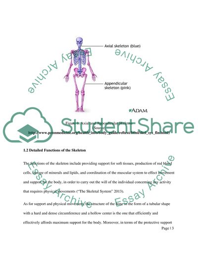
?The Skeletal System by 0 THE SKELETAL SYSTEM 1 Differences between Axial andAppendicular Skeleton The bones that make up the axial skeleton obviously differ from those that make up the appendicular skeleton. The axial skeleton is made up of skull, the hyoid bone, the auditory ossicles, the vertebral column, and the thorax, which is made up of the ribs and the sternum, or breastbone. The axial skeleton is made up of 80 bones and comprises roughly 40% of the total number of bones in the human body. Basically, the axial skeleton functions as a protective case for the internal organs of the central nervous system, which is protected by the skull and the spinal column, and the heart, which is protected by the rib cage and the breastbone (“Axial and Appendicular Skeleton” 2013). The appendicular skeleton, on the other hand, as the name implies, consists of the appendages, which include the upper limbs and the lower limbs. The upper limbs include the humerus, the radius and ulna, and the carpals and metacarpals. The lower limbs include the femur, the tibia, the fibula, the tarsals, and the metatarsals. Other components of the appendicular skeleton include the shoulder girdles, consisting of the scapulae and the collar bones, or clavicles, and the pelvic girdle, made up of the coxal or hip bones, which include the ilium, ischium and pubis. The appendicular skeleton functions for helping the various joints of the body perform better and make the body run in a smoother fashion. The appendicular skeleton too functions for movement, locomotion and the performance of any tasks done by the arms, hands, legs and feet (“Axial and Appendicular Skeleton”). Figure 1. Axial and Appendicular Skeleton (Source: http://www.pennmedicine.org/health_info/body_guide/reftext/html/skel_sys_fin.html) 1.2 Detailed Functions of the Skeleton The functions of the skeleton include providing support for soft tissues, production of red blood cells, storage of minerals and lipids, and coordination of the muscular system to effect movement and support for the body, in order to carry out the will of the individual concerning any activity that requires physical movements (“The Skeletal System” 2013). As for support and physical movement, the structure of the bone in the form of a tubular shape with a hard and dense circumference and a hollow center is the one that efficiently and effectively affords maximum support for the body. Moreover, in terms of the protective support that it gives the soft organs, the skull protects the brain; the rib cage and sternum protect the lungs and the heart; and the pelvis and the pelvic girdle provide protection for the reproductive system. In terms of movement, the bones provide anchorage for the muscles. The origin is where the muscle is fixed to a bone, and the insertion is the moving point of attachment. Finally, still in terms of the movement function, bones meet other bones at joints in order to allow varying degrees of movement (“The Skeletal System – Introduction” 2013). Another function of the skeletal system, particularly the bone marrow, is to carry out hematopoiesis, or the process by which a single type of stem cell gives rise to all types of mature red blood cells in the body. In the adult human being, this is carried out by the bone marrows of the skull, and those of the ribs, sternum, vertebra, pelvis, and the proximal ends of the thigh bones or femurs (Ownby 2002). In terms of the storage of minerals and lipids, the bone is where several metabolically active minerals are stored, especially calcium, which is the most abundant mineral in the human body. Calcium is essential in regulating the intracellular activities of muscle cells and neurons. Moreover, lipids are stored in the yellow marrow of the bone. These lipids are essential in the regulation of body heat, for providing heat, and as a structural component of cell membranes (“Osseous Tissue and Bone Structure” 2013). 1.3 Structure of Bone Tissue The biggest difference between a compact bone and a spongy bone is that the former is made up of haversian canals while the latter is made up of trabeculae. The haversian canals or osteons of the compact bone are closely packed together, and these are surrounded by numerous lamellae of the matrix. Within the matrix, there are spaces called lacunae, and it is within these that the bone cells, or osteocytes, are located. The canals that connect the lacunae to the haversian systems are known as canaliculi. The compact structure of the haversian systems creates the hardness of the compact bone as well as its appearance as a solid mass. The bone membrane is the periosteum, which contains a rich supply of blood vessels for the sustenance of the bone cells (“Structure of Bone Tissue” 2013). Figure 2. Structure of a Bone Tissue (Source: http://training.seer.cancer.gov/anatomy/skeletal/tissue.html) The spongy bone tissue is generally less dense and lighter than the compact bone. It consists of trabeculae, or plates, and bars of bone which are beside the small cavities containing the red bone marrow. The trabeculae are actually arranged in such a way that it provides maximum strength is guaranteed. Moreover, the trabeculae are flexible in case there is stress to the bone and if this stress changes (“Structure of Bone Tissue” 2013). The compact bone is found in the hard outer layer of bones, while the spongy bone makes up the bone’s internal structure. 1.4 Role and Position of Joints in the Human Skeleton One of the joints in the human body is the ball-and-socket type of joint, which may be found in the connection between the head of the femur and the acetabulum of the hipbone. This joint particularly allows freedom of rotation in all planes, and such rotation includes back-and-forth movements (“Synovial Joint Movements” 2013). Source: http://faculty.stcc.edu/AandP/AP/AP1pages/Units5to9/joints/synovial.htm Another type of joint in the human body, the pivot joint, is the type where one bone spins around another usually in two directions. Such type of joint is found in the first two cervical vertebrae as well as in the elbow, which gives it its twisting motion (“Synovial Joint Movements” 2013). Source: http://faculty.stcc.edu/AandP/AP/AP1pages/Units5to9/joints/synovial.htm Another type of human joint – the hinge joint – allows movement in only one plane, without sliding from side to side or twisting. The connection between the humerus and the ulna forms a hinge joint, which allows the movement of the arm as when the palm touches the shoulder, or when one carries dumbbells (“Synovial Joint Movements” 2013). Source: http://faculty.stcc.edu/AandP/AP/AP1pages/Units5to9/joints/synovial.htm The gliding joint, on the other hand, is a type of joint which gives the body a huge degree of flexibility such as rotation and movement in multiple directions, but not a great distance. One example of a gliding joint is where the carpals meet (“Synovial Joint Movements” 2013). Source: http://faculty.stcc.edu/AandP/AP/AP1pages/Units5to9/joints/synovial.htm The saddle joint is another kind of human joint where both bones have odd shapes but they totally complement each other. Both bones have concavities and convexities that somehow fit at every portion. An example of a saddle joint is the joint formed by the trapezium and the first metacarpal (“Synovial Joint Movements” 2013). Source: http://faculty.stcc.edu/AandP/AP/AP1pages/Units5to9/joints/synovial.htm The condyloid joint is another type of human joint where the two bones can move in many directions together with each other but they cannot rotate. It actually derives its name from condyle, or the curved process that perfectly fits into the fossa of another bone for articulation. This joint is characteristic of the joint between the mandible and the temporal bone. The lower jaw can be moved in several directions but certainly it cannot be rotated (“Synovial Joint Movements” 2013). Source: http://faculty.stcc.edu/AandP/AP/AP1pages/Units5to9/joints/synovial.htm Fibrous joints, or synarthroidal joints, are actually held together by a single ligament, and are generally immovable. Two examples are the joint between the teeth and their bony sockets, and the joints of the bones of the skull (“Types of Joint” 2013). Source: http://www.zoology.ubc.ca/~biomania/tutorial/bonejt/anc01.htm Another type of joint, the cartilaginous joint, is present in the connections between articulating bones and is definitely made up of cartilage. Cartilaginous joints are found between the vertebrae (“Types of Joint” 2013). Source: http://www.medicalook.com/human_anatomy/organs/Cartilaginous_joints.html 1.5 Three Types of Muscle Tissue in the Human Body The first and most common type of muscle tissue, and the one that is responsible for physical movement, is the skeletal muscle. The skeletal muscle is attached to the skeleton and is responsible for the individual’s posture and motor movements. However, some form of skeletal muscle is found in the soft tissues like the tongue, the diaphragm, the pharynx and the upper part of the esophagus. The skeletal muscles are generally characterized by striations formed by the actin and myosin filaments that make up the sarcomere, which is the contractile unit of the muscle. The skeletal muscle is basically voluntary and is made up of several nuclei located peripherally. Skeletal muscles are also uniformly thick throughout their length and occur in spindle shapes without branches. Skeletal muscles make up the bulk of the muscles of the body such as the triceps, the biceps and the gluteus muscles (“Muscle Tissue” 2013). The second type of muscle tissue in the human body is the smooth or visceral muscle, which is unstriated in appearance and involuntary in terms of function. The smooth muscle is found in the internal part of the internal organs and inside arteries, veins, capillaries, arterioles and venules. The lack of striations is made possible by a particular arrangement of the actin and myosin filaments. One characteristic of smooth muscle fibers is the ability to secrete elastin and collagen, which are both connective tissue matrix. Since the contraction of smooth muscles is involuntary, such muscle type responds not to human will but to autonomic and hormonal stimuli, and its contractions are slow and prolonged (“Muscle Tissue” 2013). The cardiac muscle is the third type of muscle, and is found only in the heart, especially at the base of the venae cavae. It is also an involuntary muscle but autonomic and hormonal stimuli are responsible for regulating this type of muscle. Moreover, the presence of actin and myosin filaments in the cardiac muscle, which also form sarcomeres like the skeletal muscles, accounts for the striations among this type of cell. Perhaps, the peculiar characteristic of cardiac muscles is that they are branching and that they consist of intercalated discs. Intercalated discs contain several types of cell junctions that facilitate intercellular electrical impulses. Moreover, the cardiac muscle contains Purkinje fibers, or fibers which are larger than cardiac fibers and are more efficient at conducting electrical impulses (“Muscle Tissue” 2013). 2.1 Three Diseases/Disorders of the Skeleton One bone disease that is usually seen in aging women is osteoporosis. This is characterized by the weakening of the bones and their gradual development into brittleness. It is normally brought about by old age, having a small and thin physical structure, having a low bone density, and having a family history of the disease (“Osteoporosis” 2013). Although the early stage is silent, osteoporosis gradually develops into symptoms of pain in the spine even in the absence of an injury. There can also be a loss of up to 6 inches of height and the development of a stooping posture, which is known as kyphosis. Modes of treatment for osteoporosis include medication like estrogens, teriparatide, biphosphonates or calcitonin. This should be complemented with exercise, diet, and a supervised intake of calcium and vitamin D (“Osteoporosis Overview” 2013). Osteoarthritis, or degenerative joint disease, is more popularly known as arthritis and it is its most common form. It is characterized by swelling and pain as well as reduced motion in the area of the affected joint or joints especially the spine, knees, hands and hips. It is usually caused by an overweight structure, old age, or a joint injury (“Osteoarthritis” 2013). Unfortunately, osteoarthritis cannot be treated and will just get worse over time. However, surgery and some forms of painkillers can remove the pain for proper pain management (“Osteoarthritis Overview” 2013). A third disease of the skeletal system is osteomalacia, or more popularly known as the softening of the bones caused by the inability of the body to break down and use up vitamin D. The causes of the disease include a lack of vitamin D in the diet, the occurrence of rickets, a lack of exposure to sunlight, and an intestinal condition causing malabsorption of vitamin D (“Osteomalacia” 2013). In order to treat osteomalacia, what is needed is supplementation with calcium, vitamin D and phosphorus usually taken orally. Large doses of calcium and vitamin D are also recommended for those with absorption problems in their intestine (“Osteomalacia Overview” 2013). REFERENCES “Axial and Appendicular Skeleton.” (2013). Ivy Rose, viewed 9 June 2013, “Muscle Tissue.” University of Ottawa, viewed 20 June 2013, “Osseous Tissue and Bone Structure.” (2013). Harford Community College, viewed 16 June 2013, “Osteoarthritis Overview.” The New York Times, viewed 21 June 2013, “Osteoarthritis.” National Institutes of Health, viewed 18 June 2013, “Osteomalacia Overview.” The New York Times, viewed 20 June 2013, “Osteomalacia.” National Institutes of Health, viewed 18 June 2013, “Osteoporosis Overview.” The New York Times, viewed 21 June 2013, “Osteoporosis.” National Institutes of Health, viewed 18 June 2013, Ownby, C. (2002). “Hematopoiesis.” Oklahoma State University, viewed 16 June 2013, “Structure of Bone Tissue.” (2013). SEER Training Modules. National Cancer Institute, viewed 18 June 2013, “Synovial Joint Movements.” (2013). Springfield Technical Community College, viewed 18 June 2013, “The Skeletal System - Introduction.” (2013). Union County College, viewed 18 June 2013, “The Skeletal System.” (2013). Pearson Education, viewed 11 June 2013, “Types of Joint.” Teach PE, viewed 20 June 2013, Read More
Cite this document
  • APA
  • MLA
  • CHICAGO
(“The Skeletal System Assignment Example | Topics and Well Written Essays - 2000 words”, n.d.)
Retrieved from https://studentshare.org/nursing/1479911-the-skeletal-system
(The Skeletal System Assignment Example | Topics and Well Written Essays - 2000 Words)
https://studentshare.org/nursing/1479911-the-skeletal-system.
“The Skeletal System Assignment Example | Topics and Well Written Essays - 2000 Words”, n.d. https://studentshare.org/nursing/1479911-the-skeletal-system.
  • Cited: 0 times

CHECK THESE SAMPLES OF The Skeletal System

Analysis of the 6 Categories of Leukocytes

The Skeletal System provides the body with its basic framework, as well as the structure.... Word documents are acceptable by the system, uploaded documents can not be graded automatically and thus create major problems.... BIOL 301, Spring 2013: Answer Sheet Instructor: Dr....
4 Pages (1000 words) Essay

Structure of the Bone

The Skeletal System is one of the most important systems of the body.... The Skeletal System of the human body comprises of 206 bones.... The Skeletal System also has many structural as well as physiological roles that it performs (Alcamo and Krumhardt 2004; Plowman and Smith 2007).... These muscles are in turn regulated by the impulses from the central nervous system.... hellip; skeletal muscles are attached to these bones which assist in their movement....
3 Pages (750 words) Essay

The Skeletal System of a Human

This paper "The Skeletal System of a Human" focuses on the fact that there are 206 bones in the body and all of those combined to give the framework is known as The Skeletal System.... Bones are not the only important part of The Skeletal System; the other important feature is the joints.... nbsp;… The system performs five main functions....
1 Pages (250 words) Essay

The Human Anatomy and Possible Threats

The main systems of the human body are the cardiovascular system, the digestive system, the endocrine system, the urinary system, the immune system, the muscular system, the nervous system, the reproductive system, and the respiratory system (Schlossberg and Zuidema, 2007).... The circulatory system is responsible for the circulation of blood and lymph around the body (Swearingen, 2012)....
12 Pages (3000 words) Essay

Skeletal System in the Human Body

What critical functions are performed by The Skeletal System?... The Skeletal System provides the mechanical levers for certain groups of muscles whose contraction moves the bodyHousing bone marrow which produces red blood cellsStoring calcium and phosphorus.... The paper "skeletal system in the Human Body " describes that skeletal muscles are more rapid in contracting than cardiac and smooth muscles.... he cartilages are said to be avascular there is no canal system or blood vessel penetrating the cartilage structureBriefly describe the diarthroses and synarthroses types of joint....
2 Pages (500 words) Assignment

Mitochondria as an Essential Part of Functioning of the Organelles in the Endomembrane

The Skeletal System is composed of cartilage, bones and ligaments.... The Skeletal System protects and supports muscles that are attached to bones as well as the body; provides calcium storage and is also a site where blood cells are formed (John, 2010).... There are several organ systems that include; integumentary system made up of skin, nails, hair and sweat glands which is responsible for covering and protecting the body.... he muscular system on the other hand comprises of cardiac muscle, skeletal muscle and smooth muscle....
4 Pages (1000 words) Essay

Muscular and Skeletal system Worksheet

Diet – Diet affects bone health by allowing nutrients required to maintain the strength of bones to reach The Skeletal System.... Diet – Diet affects bone health by allowing nutrients required to maintain the strength of bones to reach The Skeletal System.... Exercise – Regularly working out rouses the renovation of The Skeletal System.... Proper cells include Muscular and skeletal system Worksheet Muscular and skeletal system Worksheet Questions List two factors that keep bones healthy....
1 Pages (250 words) Assignment

How Does Thalassemia Affect the Skeletal System

Thalassemia is a Greek word for sea blood that is, the place of origin of where the disorder was first discovered among people living around the Mediterranean Sea (Ortner, 2003).... To date, the cases of Thalassemia are still high in these regions, and the genes carrying the… Thalassemia affects both men and women and occurs in an estimated 1 of every 100000 individuals worldwide....
6 Pages (1500 words) Essay
sponsored ads
We use cookies to create the best experience for you. Keep on browsing if you are OK with that, or find out how to manage cookies.
Contact Us