StudentShare

Our website is a unique platform where students can share their papers in a matter of giving an example of the work to be done. If you find papers
matching your topic, you may use them only as an example of work. This is 100% legal. You may not submit downloaded papers as your own, that is cheating. Also you
should remember, that this work was alredy submitted once by a student who originally wrote it.
✕
Free
Practical Experience of Routine Staining in Histopathology Lab - Term Paper Example
Summary
The paper "Practical Experience of Routine Staining in Histopathology Lab" tells that the method achieves this by clearly straining cell structure. Some of the cell components strained is the nucleus, cytoplasm, organelles, and extracellular components…
- Subject: Chemistry
- Type: Term Paper
- Level: Undergraduate
- Pages: 4 (1000 words)
- Downloads: 0
- Author: kesslercarley
Extract of sample "Practical Experience of Routine Staining in Histopathology Lab"
Practical 2. Hematoxylin & eosin staining, and frozen tissue embedding s Most of the organism Fresh tissues specimens are colorless. They provide very little information, and it is hard to differentiate between tissues. This experiment is objective is to help the student gain practical experience of routine staining in histopathology lab and to allow them to view how frozen tissue are prepared. From the result of the experiment, student was able to identify that embedding and staining are some of the methods used to produce the frozen section. They also identified that H & E is the most common staining method. According to the experiment result, the method is routinely used in histopathology lab because it provides enough detail for tissues and cell diagnostic. Using the microscope student observed and labeled several components of the uterus tissue like Endometrial glands, Stroma, Myometrium. It was also observed that the cell nucleus turned red and cytoplasm pink after H&E staining.
Introduction
Normally most of the cells are transparent, with little or no pigment. Even red blood cell which has hemoglobin appears almost colorless, unless there are packed in big masses (Mula, 2009). Straining make the cell components visible, this enable comparing of cells and tissues. Thus, the main purpose of training is to make cell structures easier to see. For diagnostic purpose, tissues must undergo some alteration for viewing under microscope (Brown, 2009). Fixation, embedding, slicing and staining are some of the basic step for tissue preparation.
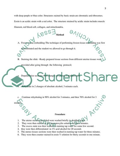
In the histopathology lab, the term routine staining refers to H& E (hexactoxylin and eosin) that is regularly used with all tissue specimens to identify the various structures. Routine H & E, staining plays a critical role in cell and tissue diagnostic. Hematoxylin is a basic strain with deep purple or blue color. Structures stained by basic strain are chromatic and ribosomes. Eosin is an acidic strain with a red color. The structure strained by acidic strain includes muscle filament, red blood cell, collagen, and mitochondria.
Method
A. Frozen tissue embedding-The technique of performing frozen tissue embedding was first demonstrated and the student we allowed to go through it.
B. Staining the slide: -Ready prepared tissue sections from different uterine tissues were provided after going through the following protocol;
a. Deparaffinize sections; two changes of xylene, 5 minutes each.
b. Rehydrate in 2 changes of absolute alcohol; 3 minutes each.
c. Continue rehydrating in 90% alcohol for 3 minutes, and then 70% alcohol for 3
minutes.
Procedure
1. The uterus sections provided were washed briefly in distilled water.
2. They were then stained in gill haematoxylin solution for three minutes.
3. The excess stain was then washed in running tap water for some few second.
4. they were then differentiated in 1% acid alcohol for 20 seconds
5. The uterus tissues sections were then washed in running tap water for three minutes.
6. They were then counter stained in eosin Y solution for thirty seconds to one minute.
7. The section was then Dehydrated through 70% alcohol for two minutes, 90% alcohol for two minutes and two changes of absolute alcohol three minutes each.
8. then they were cleared in two changes of xylene, for two minutes each.
9. they were then mounted with xylene-based mounting medium
3. Light microscopy on the specimen - The light microscope was set and used it to examine the slide. The nucleic was observed to be blue and cytoplasm red. The images observed under the microscope were drawn, and the following components were labeled: Endometrial glands,S troma and Myometrium.
Results
1. Endometrial glands are stained pink/purple
2. Stroma is stained bright pink
3. Myometruim bright red / pink
Discussion
One advantage of preparing frozen section is that, it can be prepared first as compared to other staining methods. The frozen sections are largely used to examine tissues in during surgery. For instance, during the diagnosis of aoasophagus tumors, frozen section help to define resection margins for pastroesophageal resection. Moreover the method helps to evaluate the extent of disease spread. In such a case, frozen section may aid in complete resection of the tumors cell (Kim, & Weickert, 2007).
Each step introduces artifacts by distorting the cell natural appearance. Some artifacts are avoidable while others are unavoidable. Among the most common artifacts are wrinkles and scratches. Scratched are caused by dirt on the cutting edge. The procedure should be carried out with care to avoid unintended artifact since they can result to wrong diagnostic. Unintended artifact can be minimized by optimal procedure.
From the examination of the uterus tissue sections, some endometrial gland contained narrow lumen. This was an indication of folical phase. The luteal phase was also observed (Weickert, 2009). At the beginning of the phase, progesterone stimulates secretion osecretef mucus and glycogen by the endometrial glands. The large lumens are, therefore, as a result of increased secretary activity.
There were no signs of fibroid or endometriosis observed.
Conclusion
H & E is the most often used staining method in the histology lab for tissue diagnostic. H&E is mostly used as it provides pathologist a very detailed view of the tissue (Kiernan, 2009). The method achieves this by clearly straining cell structure. Some of the cell components strained is nucleus, cytoplasm, organelles, and extra cellular components. The information from HE& E is sufficient to allow diagnosis of disease and to show abnormalities. It can also show particular indicators of abnormalities such as nuclear changes common in cancerous cells.
Errors are occurring during the staining lead to false diagnostic. Mismatching is one of the common errors that occur during staining. It may result from, mixing of specimens. To avoid mismatching, it is important to clearly label all specimens from the patients.
References
Brown RW 2009 ‘Histologic Preparations: Common Problems and Their Solutions. College of American Pathologists, Northfield IL
Kiernan JA.2009 Histology FAQ Staining, Histochemistry and Histotechnology. Accessed October 5, 2009 at: http://www.ihcworld.com/_faq/ histology-faq/misc/m6.htm.
Kim YJ., &, Weickert J 2007, ‘Fully automated segmentation and morphometrical analysis of muscle fiber images. Vol 17, no ,71, pp. 8–15.
Mula J, L 2009, ‘Automated image analysis of skeletal muscle fiber cross-sectional area.’vol. 35, np 34, pp. 148–155
Weickert J 2009, ‘Coherence-enhancing diffusion filtering’ vol. 6 , no. 31, pp. 111–127.
Read
More
sponsored ads
Save Your Time for More Important Things
Let us write or edit the term paper on your topic
"Practical Experience of Routine Staining in Histopathology Lab"
with a personal 20% discount.
GRAB THE BEST PAPER
