StudentShare
Contact Us
Sign In / Sign Up for FREE
Search
Go to advanced search...
Free

Scientific Principles of Magnetic Resonance Imaging - Essay Example

Cite this document
Summary
This paper 'Scientific Principles of Magnetic Resonance Imaging' tells us that Magnetic resonance imaging and ultrasound are both radiographic imaging techniques that are commonly known in terms of safety, mainly for their non-use of ionizing radiation common in other diagnostic procedures like x-ray, computerized tomography…
Download full paper File format: .doc, available for editing
GRAB THE BEST PAPER93.7% of users find it useful
Scientific Principles of Magnetic Resonance Imaging
Read Text Preview

Extract of sample "Scientific Principles of Magnetic Resonance Imaging"

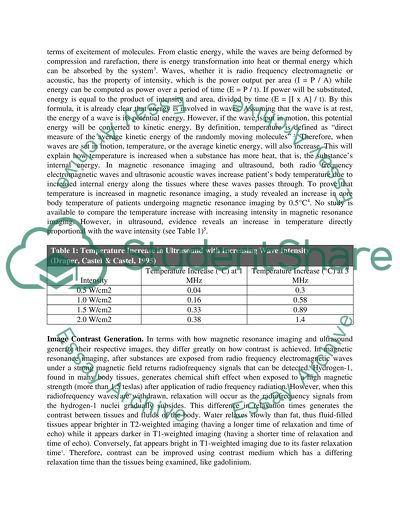
?Scientific Principles of Magnetic Resonance Imaging (MRI) and Ultrasound: A Comparative Essay Magnetic resonance imaging (MRI) and ultrasound are both radiographic imaging techniques which are commonly known in terms of safety, mainly for its non-use of ionizing radiation common in other diagnostic procedures like x-ray and computerized tomography. It is a common knowledge that ultrasound is cheaper, more portable, more rapidly available in emergency settings and generates real-time images, while magnetic resonance imaging provides greater detail, penetration to hard-to-penetrate areas in ultrasound, the dependence on magnetism and more sophisticated and expensive equipment. However, there are similarities and differences of these radiographic techniques in terms of scientific principles behind these, by which this paper intends to explore. Transmission and Reception of Waves. Magnetic resonance imaging and ultrasound shares their similarity in their ability to transmit their respective waves, electromagnetic and acoustic waves respectively, while receiving the signals induced by these transmitted waves. In magnetic resonance imaging, transmit coils emit radio waves to the patient, calibrated at the precessional frequency or Larmor frequency of hydrogen-1 at 42.6 MHz under a magnetic field strength of 1.5 tesla or above, generates a strong magnetic signal within the hydrogen-1 nucleus which can be detected by the receiver coils of the device1. On the other hand, in ultrasound, ultrasonic acoustic waves come from the source transducer as a result of a piezoelectric effect (conversion of electrical to sound energy), and solid objects along the watery body tissues causes an echo, which will be detected by the receiving transducer using the same piezoelectric effect (conversion of sound energy to electrical energy)2. Type of Waves. There are differences between electromagnetic waves emitted by magnetic resonance imaging and the acoustic waves of ultrasound imaging, mainly on the speed of wave travel (electromagnetic waves are faster than acoustic waves), ability to travel in an empty space (acoustic waves cannot travel in a vacuum while electromagnetic waves can), and the type of waves traveling along fluid (longitudinal and transverse in electromagnetic waves, while longitudinal only in acoustic waves), yet both electromagnetic and acoustic waves are similar to be having properties of frequencies, amplitude, intensity2. How these properties are used in both devices is also dissimilar (the frequency of electromagnetic waves in MRI is set to a standard 42.6 MHz setting, while the frequency in ultrasound is calibrated according to the depth of penetration)1 2. Non-use of ionizing radiation. Both magnetic resonance imaging and ultrasound do not utilize ionizing radiation present in x-ray, computerized tomography and fluoroscopy. In magnetic resonance imaging, the electromagnetic wave frequency of 42.6 MHz is very low to cause ionization of molecules, while acoustic waves are not a type of radiation which causes ionization of molecules even in high frequencies2. Body Temperature Elevation. Both magnetic resonance imaging and ultrasound produces heat. Heat is a form if energy, which can be appreciated in thermodynamics as “internal energy” in terms of excitement of molecules. From elastic energy, while the waves are being deformed by compression and rarefaction, there is energy transformation into heat or thermal energy which can be absorbed by the system3. Waves, whether it is radio frequency electromagnetic or acoustic, has the property of intensity, which is the power output per area (I = P / A) while energy can be computed as power over a period of time (E = P / t). If power will be substituted, energy is equal to the product of intensity and area, divided by time (E = [I x A] / t). By this formula, it is already clear that energy is involved in waves. Assuming that the wave is at rest, the energy of a wave is its potential energy. However, if the wave is put in motion, this potential energy will be converted to kinetic energy. By definition, temperature is defined as “direct measure of the average kinetic energy of the randomly moving molecules” 2. Therefore, when waves are set in motion, temperature, or the average kinetic energy, will also increase. This will explain how temperature is increased when a substance has more heat, that is, the substance’s internal energy. In magnetic resonance imaging and ultrasound, both radio frequency electromagnetic waves and ultrasonic acoustic waves increase patient’s body temperature due to increased internal energy along the tissues where these waves passes through. To prove that temperature is increased in magnetic resonance imaging, a study revealed an increase in core body temperature of patients undergoing magnetic resonance imaging by 0.5°C4. No study is available to compare the temperature increase with increasing intensity in magnetic resonance imaging. However, in ultrasound, evidence reveals an increase in temperature directly proportional with the wave intensity (see Table 1)5. Table 1: Temperature Increase in Ultrasound with Increasing Wave Intensity (Draper, Castel & Castel, 1995) Intensity Temperature Increase (°C) at 1 MHz Temperature Increase (°C) at 3 MHz 0.5 W/cm2 0.04 0.3 1.0 W/cm2 0.16 0.58 1.5 W/cm2 0.33 0.89 2.0 W/cm2 0.38 1.4 Image Contrast Generation. In terms with how magnetic resonance imaging and ultrasound generate their respective images, they differ greatly on how contrast is achieved. In magnetic resonance imaging, after substances are exposed from radio frequency electromagnetic waves under a strong magnetic field returns radiofrequency signals that can be detected. Hydrogen-1, found in many body tissues, generates chemical shift effect when exposed to a high magnetic strength (more than 1.5 teslas) after application of radio frequency radiation. However, when this radiofrequency waves are withdrawn, relaxation will occur as the radiofrequency signals from the hydrogen-1 nuclei gradually subsides. This difference in relaxation times generates the contrast between tissues and fluids of the body. Water relaxes slowly than fat, thus fluid-filled tissues appear brighter in T2-weighted imaging (having a longer time of relaxation and time of echo) while it appears darker in T1-weighted imaging (having a shorter time of relaxation and time of echo). Conversely, fat appears bright in T1-weighted imaging due to its faster relaxation time1. Therefore, contrast can be improved using contrast medium which has a differing relaxation time than the tissues being examined, like gadolinium. However, in ultrasound, instead of radio frequency electromagnetic signals from ferromagnetic substances like hydrogen-1, contrast is achieved using the differences in echoes from solid objects, measuring the time it takes for the echo to be received and the intensity of the sound waves. Both the time of echo and the intensity of the acoustic wave decrease as the tissue is farther from the transducer. The frequency of the acoustic wave is adjusted according to the depth of penetration of the scan (higher frequency on the more superficial organs like optical scan, while lower frequency on more visceral organs like the abdomen). The pattern of the differences in echo intensity and time of echo generates the varying contrast in the ultrasound display2. To enable enhanced visualization of body tissues, the speed of sound in ultrasound machines is set to 1540 m/s since the speed of sound in most of the fluid-filed body tissues are close to this setting6. However, though ultrasound can travel faster in bones (4080 m/s), the acoustic waves has difficulty traveling along bones due to it’s non-fluid filled composition 7. Dense fluid-filled body tissues appear brighter (at the desired frequency level), while bones are not visible. This difficulty in penetrating through bones is shared by magnetic resonance imaging as both depends on the fluid content of tissues. Another difficulty in ultrasound imaging, but not true for magnetic resonance imaging, is on traveling through air for the reason that the speed of sound in air is slower than fluids and fluid-filled tissues. This is the reason why ultrasound is hindered by intestinal gas, while it can be enhanced by fluid intake8. Motion Visualization. Magnetic resonance imaging and ultrasound can visualize trends of body fluids over time, in different scientific principles, which allows the detection of blockages and obstructions. Magnetic resonance imaging uses the gradient echo imaging technique wherein signals are refocused from low flux density to higher flux density, allowing increased exposure to flowing fluid (blood)1. An example of the use of magnetic resonance imaging in detection of obstructions is in the case of arterial thrombus9. On the other hand, ultrasound uses the Doppler effect to measure the velocity of the fluid as there are differences in frequencies of echo from the reflecting acoustic waves, and it is possible to visualize the motion by filtration of frequencies from stable substances / tissues. In case of blockages, the blood flow along the obstruction appears faster than the normal blood vessel2. An example of detection of obstruction using Doppler ultrasound is in cases of atherosclerosis10. Conclusion This paper compared and contrasted the scientific principles behind magnetic resonance imaging from ultrasound. These radiographic procedures are similar in terms of how their respective waves are transmitted and how the echo waves are received. They do not use ionizing radiation but able to increase body temperature (and heat). They differ mainly on how image is rendered. Both techniques can visualize obstructions or blockages, but in a different way. This paper briefly mentioned other similarities and differences in the introduction since the scope of this essay is on scientific principles. Read More
Cite this document
  • APA
  • MLA
  • CHICAGO
(“Compare and contrast the scientific principles of Magnetic resonance Essay”, n.d.)
Retrieved from https://studentshare.org/environmental-studies/1412727-compare-and-contrast-the-scientific-principles-of
(Compare and Contrast the Scientific Principles of Magnetic Resonance Essay)
https://studentshare.org/environmental-studies/1412727-compare-and-contrast-the-scientific-principles-of.
“Compare and Contrast the Scientific Principles of Magnetic Resonance Essay”, n.d. https://studentshare.org/environmental-studies/1412727-compare-and-contrast-the-scientific-principles-of.
  • Cited: 0 times

CHECK THESE SAMPLES OF Scientific Principles of Magnetic Resonance Imaging

Magnetic Resonance Imaging

[Name] [Subject] [Course] [Instructor] [Date] magnetic resonance imaging magnetic resonance imaging (MRI) is a method to scan the body's internal organs mostly used in the medical facilities to cast an image of internal body systems and internal parts.... magnetic resonance imaging magnetic resonance imaging (MRI) is a method to scan the body's internal organs mostly used in the medical facilities to cast an image of internal body systems and internal parts....
3 Pages (750 words) Research Paper

Nanoprobes used to Fight Breast Cancer Tumors

To put this in proportion we can compare this to being the same as about ten times the diameter of a hydrogen atom.... 'Breast Cancer' sounds terrifying to most women.... This is due to the high incidence of the disease as well as the image it conjures up of painful treatments and awful side effects....
28 Pages (7000 words) Essay

Child Expectations during Magnetic Resonance Imaging

The scanner for mri resembles a huge box that is square shaped and with a tunnel-like hole running through the middle.... When the child has been set in a comfortable and appropriate position, the table begins to move by default into the tunnel of the mri.... Upon starting the process of mri, the child may experience sounds that are loud, knocking and thumping in nature that may stay for many minutes at times.... A parent/ guardian can come along with the child's favorite DVD so that they can watch during the process through the video goggles of the mri or even music CDs that they can listen to during the process....
7 Pages (1750 words) Essay

Pregnancy Risks & Scanning Methods - Computed Tomography, Magnetic Resonance Imaging

These methods include Computed Tomography (CT), magnetic resonance imaging (MRI) and Ultrasound (US) and they are all utilised to examine and evaluate… The purpose of this paper is to examine the possible biological risks and hazards that are associated with these three methods when imaging a pregnant patient.... This will be done by the examination of the scientific In the process recommendations of how to reduce and ameliorate these risks are critiqued and examined A computed tomography (CT) scan utilises x-rays to provide detailed pictures of structured inside the body of pregnant women (WebMD, 2013)....
8 Pages (2000 words) Essay

The Basic Principles of the Magnetic Resonance Imaging Image Production

This paper "The Basic Principles of the magnetic resonance imaging Image Production" focuses on the fact that magnetic resonance imaging is a new, revolutionary, and exciting imaging technique used in generating images of the internal structural anatomy of the body to help in the diagnosis.... RI is essentially dependent on the principles of magnetism or the ability of matter to be attracted to other matter.... nbsp;… The main component of an mri is a magnetic field which is used to stabilize the hydrogen protons and prepare them for manipulation or excitation through the introduction of electromagnetic pulses....
7 Pages (1750 words) Case Study

Basic Principles of Image Production of Magnetic Resonance Imaging

This paper "Basic Principles of Image Production of magnetic resonance imaging" focuses on the fact that MRI is one of the most preferred diagnostic tools in healthcare.... The relation between the magnetic resonance and the location makes it possible to reconstruct the image being scanned by the imaging process.... 106) discuss that mri is “based on the use of inhomogeneous or 'gradient' fields supplementing on an otherwise uniform invariant magnetic field”....
8 Pages (2000 words) Case Study

Magnetic Resonance Imaging

This paper will address the natural susceptibility of the developing neonatal brain in preterm infants, the function of magnetic resonance imaging (MRI) as an instrument in the detection of atypical growth.... magnetic resonance imaging (MRI) is a noninvasive health procedure that assists physicians in the detection and treatment of medical conditions.... The use of a more effective and accurate neuroimaging method, such as magnetic resonance imaging (MRI), is now widely preferred in the detection of slight to severe injuries of the infant's brain (Woodward et al)....
6 Pages (1500 words) Research Paper

Magnetic Resonance Imaging and Ultrasound Scans

The paper "magnetic resonance imaging and Ultrasound Scans" highlights that CT provides a system of scanning and reviewing important trends and features in the body of a pregnant woman.... hellip; Most authorities identify that ultrasound scans of foetuses have little or no connection to any known sicknesses and deformities in children (Rosene-Montella, 2009).... However, some research works conducted show a possible connection between some deformities and defects in the development of babies with ultrasound scans....
9 Pages (2250 words) Coursework
sponsored ads
We use cookies to create the best experience for you. Keep on browsing if you are OK with that, or find out how to manage cookies.
Contact Us