StudentShare
Contact Us
Sign In / Sign Up for FREE
Search
Go to advanced search...
Free

Muscle Strain and Repair - Essay Example

Cite this document
Summary
This paper "Muscle Strain and Repair" recommends stretching and strengthening exercises as part of a physical conditioning program to minimize the risk of muscle strains, protect joints in the long term by working to strengthen and condition the muscles around the joint that has been injured…
Download full paper File format: .doc, available for editing
GRAB THE BEST PAPER93.7% of users find it useful
Muscle Strain and Repair
Read Text Preview

Extract of sample "Muscle Strain and Repair"

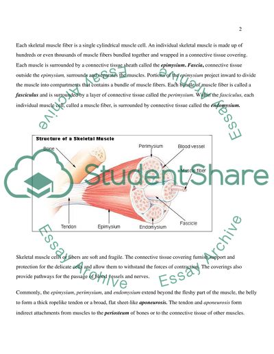
Muscle Strain and Repair A skeletal muscle is considered an organ of the muscular system. Each organ or muscle consists of skeletal muscle tissue, connective tissue, nerve tissue, and blood or vascular tissue. In addition to movement, muscle contraction fulfills other important functions in the body, such as posture, joint stability, and heat production. Posture such as sitting and standing is maintained as a result of muscle contraction. The muscular system is composed of specialized cells called muscle fibers. Their predominant function is contractibility. The muscle fibers are the basic contractile units of skeletal muscles. They are individually surrounded by a connective tissue layer and grouped into bundles to form a skeletal muscle. As well as being rich in connective tissue, skeletal muscles are highly vascularized to provide essential nutrients for muscle function. Muscles attached to bones or internal organs and blood vessels, are responsible for movement. Movement in the body is the result of muscle contraction. The integrated action of joints, bones, and skeletal muscles produces movements such as walking and running. Skeletal muscles also produce the subtle movements that result in various facial expressions, eye movements, and respiration. The skeletal muscles continually make fine adjustments that hold the body in stationary positions. The tendons of many muscles extend over joints and in this way contribute to joint stability. This is particularly evident in the knee and shoulder joints, where muscle tendons are a major factor in stabilizing the joint. Heat production, to maintain body temperature, is an important by-product of muscle metabolism. Nearly 85 percent of the heat produced in the body is the result of muscle contraction. Structure of a Skeletal muscle Skeletal muscles vary considerably in size, shape, and arrangement of fibers. They range from extremely tiny strands such as the stapedium muscle of the middle ear to large masses such as the muscles of the thigh. Some skeletal muscles are broad and some narrow in shape. In some muscles the fibers are parallel to the long axis of the muscle, in some they converge to a narrow attachment, and in some they are oblique. Each skeletal muscle fiber is a single cylindrical muscle cell. An individual skeletal muscle is made up of hundreds or even thousands of muscle fibers bundled together and wrapped in a connective tissue covering. Each muscle is surrounded by a connective tissue sheath called the epimysium. Fascia, connective tissue outside the epimysium, surrounds and separates the muscles. Portions of the epimysium project inward to divide the muscle into compartments that contains a bundle of muscle fibers. Each bundle of muscle fiber is called a fasciculus and is surrounded by a layer of connective tissue called the perimysium. Within the fasciculus, each individual muscle cell, called a muscle fiber, is surrounded by connective tissue called the endomysium. Skeletal muscle cells or fibers are soft and fragile. The connective tissue covering furnish support and protection for the delicate cells and allow them to withstand the forces of contraction. The coverings also provide pathways for the passage of blood vessels and nerves. Commonly, the epimysium, perimysium, and endomysium extend beyond the fleshy part of the muscle, the belly to form a thick ropelike tendon or a broad, flat sheet-like aponeurosis. The tendon and aponeurosis form indirect attachments from muscles to the periosteum of bones or to the connective tissue of other muscles. Typically a muscle spans a joint and is attached to bones by tendons at both ends. One of the bones remains relatively fixed or stable while the other end moves as a result of muscle contraction. Skeletal muscles have an abundant supply of blood vessels and nerves. This is directly related to the primary function of skeletal muscle, contraction. Before a skeletal muscle fiber contract, it receives an impulse from a nerve cell. Generally, an artery and at least one vein accompany each nerve that penetrates the epimysium of a skeletal muscle. Branches of the nerve and blood vessels follow the connective tissue components of the muscle of a nerve cell and with one or more minute blood vessels called capillaries. Muscle Injury: A strain is a stretching or tearing of muscle. This type of injury often occurs when muscles suddenly and powerfully contract or when a muscle stretches unusually far. This is called an acute strain and overuse of certain muscles over time lead to a chronic strain. People commonly call muscle strains "pulled" muscles. Hamstring and back injuries are among the most common strains. Treatment depends on the severity of the injury. Mild strain is when pain and stiffness that occur with movement last a few days. Moderate strain is partial muscle tears that result in more extensive pain, swelling and bruising and the pain last one to three weeks. Severe strain is when the muscle is torn apart or ruptured. There is significant bleeding, swelling and bruising around the muscle. Muscle may not function at all, and surgical repair is required if the muscle has torn away completely from the bone. Causes of muscle strain: Strains occur commonly and most result in minor injuries. A muscle becomes strained or pulled or may even tear when it stretches unusually far or abruptly. This type of injury, an acute strain often occurs when muscles suddenly and powerfully contract. A muscle strain may occur when one slip on ice, run, jump, throw, lift a heavy object or lift in an awkward position. A chronic strain results from prolonged, repetitive movement of a muscle. One may sprain knee or ankle when walking or exercising on an uneven surface. A sprain also may occur when one land awkwardly, either at the end of a jump or while pivoting during an athletic activity. Poor conditioning: Lack of conditioning leave muscles weak and more likely to sustain injury. Poor technique: The way when one land from a jump for example, when skiing or practicing martial arts may affect risk of injury to a ligament in knee called the anterior cruciate ligament ACL. Landing with an inward rotation at the knee "knock-kneed" position predispose one to an ACL sprain. Fatigue: Tired muscles are less likely to provide good support for the joints. When one is tired, one is more likely to succumb to forces that could stress a joint or overextend a muscle. Improper stretching and warm-up: Properly warming up and stretching before vigorous physical activity loosens the muscles and increases joint range of motion, making the muscles less tight and less prone to trauma and tears. Repair and Healing of Muscles: Skeletal muscle is a stable tissue with very little turnover of nuclei. However, upon injury, skeletal muscle has the remarkable ability to initiate a rapid and extensive repair process preventing the loss of muscle mass. Skeletal muscle repair is a highly synchronized process involving the activation of various cellular responses. The initial phase of muscle repair is characterized by necrosis of the damaged tissue and activation of an inflammatory response. This phase is rapidly followed by activation of myogenic cells to proliferate, differentiate, and fuse leading to new myofiber formation and reconstitution of a functional contractile apparatus. Muscle satellite cell activation resembles embryonic myogenesis in several ways including the de novo induction of the myogenic regulatory factors. Skeletal muscle regeneration is a highly orchestrated process that involves the activation of adult muscle satellite cells to proliferate and differentiate. Activation of satellite cells requires the timely, controlled up regulation of muscle transcription factors and muscle specific genes. This process is regulated through mechanisms involving cell-cell and cell matrix interactions as well as extra cellular secreted factors. (Chen G, Quinn L S, 1992) Muscle injuries have been shown to cause the release of biologically active molecules into the extra cellular space. (Bischoff R, 1986) The healing process, (Almekinders LC, Gilbert JA, 1986) include an inflammatory response, parenchymal cell proliferation and migration,neovascularisation, and extra cellular matrix deposition. It is generally recognised that healing is divided into three temporal phases, which overlap each other but are distinct in terms oftime after injury. The relative importance of each phase depends on the type, location and severity of the injury. (Slavin, 1999) The healing process is one of repair, where damaged tissue is restored by the formation of connective tissue and re-growth of epithelium. (Tortora and Gradowski, 1996) Skeletal Muscle Regeneration: Skeletal muscle is a stable tissue with little turnover of nuclei (Decary S et al, 1997) Minor lesions inflicted by day-to-day wear and tear elicit only a slow turnover of its constituent multinucleated muscle fibers. It is estimated that in a normal adult muscle, no more than 1-2% of myonuclei are replaced every week (Schmalbruch H, Lewis DM, 2000) Skeletal muscle has the ability to complete a rapid and extensive regeneration in response to severe damage. Whether the muscle injury is inflicted by a direct trauma i.e., extensive physical activity and especially resistance training or innate genetic defects, muscle regeneration is characterized by two phases: Degenerative phase and Regenerative phase The initial event of muscle degeneration is necrosis of the muscle fibers. This event is generally triggered by disruption of the myofiber sarcolemma resulting in increased myofiber permeability. The primary components of skeletal muscles are the myofibers grouped in bundles within the perimysium. Skeletal muscles are highly vascularized to provide essential nutrients for muscle function. Myofibers are heterogeneous with respect to their contractile properties, ranging from slow and oxidative to fast and glycolytic types. The proportion of each fiber type within a muscle determines its overall contractile property. The adult skeletal muscle contains a population of quiescent muscle satellite cells. Muscle satellite cells are closely associated with myofibers, located within the same basal lamina. Muscle satellite cell nuclei are distinguished from myonuclei by their abundant heterochromatin reflecting their mitotic quiescence. Muscle satellite cells are present on myofibers isolated by mild enzymatic digestion and are characterized by their high levels of Pax7expression. (Bendall AJ, 1999) Inflammatory response: Skeletal muscle repair process is characterized by a degenerative phase followed by a regenerative phase. Injury to the tibialis anterior muscle by Cardio-toxin CTX injection resulted in the rapid necrosis of myofibers and the activation of an inflammatory response leading to the loss of muscle architecture. Myofiber regeneration is characterized by the activation of myogenic cells to proliferate, differentiate and fuse to necrotic fibers for repair and to each other for new fiber formation. Regenerating fibers are characterized by their small caliber and their centrally located myonuclei. The early phase of muscle injury is usually accompanied by the activation of mono-nucleated cells, principally inflammatory cells and myogenic cells. (Tidball J G, 1995) Present reports suggest that factors released by the injured muscle activate inflammatory cells residing within the muscle, which in turn provide the chemotactic signals to circulating inflammatory cells. (Rappolee DA, Werb Z, 1992) Neutrophils are the first inflammatory cells to invade the injured muscle, (Fielding RA et al, 1993) with a significant increase in their number being observed as early as 1-6 h after myotoxin or exercise induced muscle damage. (Orimo S et al, 1991) After neutrophil infiltration and 48 h post injury, macrophages become the predominant inflammatory cell type within the site of injury. (Tidball J G, 1995) Macrophages infiltrate the injured site to phagocytose cellular debris and affect other aspects of muscle regeneration by activating myogenic cells (Lescaudron L et al, 1999) A systemic factor capable of inducing an inflammatory response throughout the body is released following muscle damage (Fehr HG, Lotzerich H, Michna H, 1988) Thus muscle fiber necrosis and increased number of non muscle mono-nucleate cells within the damaged site are the main histopathological characteristics of the early event following muscle injury. Muscle degeneration is followed by the activation of a muscle repair process. Cellular proliferation is an important event necessary for muscle regeneration. The expansion of myogenic cells provides a sufficient source of new myonuclei for muscle repair (Grounds MD et al, 2002) Numerous nuclear radio-labeling experiments have demonstrated the contribution of dividing myogenic cells to regenerating myofibers, and it is well accepted that following the myogenic proliferation phase, new muscle fibers are formed much as during bona fide embryonic myogenesis. (Hawke TJ, Garry DJ, 2001) Myogenic cells differentiate and fuse to existing damaged fibers for repair and to one another for new myofiber formation (Darr KC, Schultz E, 1987) Cell fusion is not diffused during regeneration but rather focal to the site of injury. (Blaveri K et al, 1999) Fiber splitting or branching is also a characteristic feature of muscle regeneration and is probably due to the incomplete fusion of fibers regenerating within the same basal lamina (Bourke DL, Ontell M, 1984) Fiber splitting is commonly observed in muscles from patients suffering neuromuscular diseases, in hypertrophied muscles, and in aging mouse muscles, all of which are associated with abnormal regenerative capacity. (Bockhold KJ et al, 1998) Muscle Satellite cells in Muscle repair: The activation of satellite cells upon muscle injury resulting from mechanical trauma, direct injury to the muscle and in the course of a disease is well characterized (Campion DR, 1984) Moreover, when transplanted into regenerating muscle, cultured satellite cells contribute to new myofiber formation as well as to reconstitution of satellite cell population for later rounds of regeneration (Gross JG, Morgan JE, 1999) Activation of Muscle Satellite Cell Upon Injury: In the course of muscle regeneration, satellite cells first exit their normal quiescent state to start proliferating. After several rounds of proliferation, the majority of the satellite cells differentiates and fuse to form new myofibers and to repair damaged one. Satellite cell activation is not restricted to the damaged site. Damage at one end of a muscle fiber activates satellite cells all along this fiber leading to the proliferation and migration of the satellite cells to the regeneration site. (Schultz E, Jaryszak DL, Valliere CR, 1985) However, recruitment of satellite cells from adjacent muscles is seldom observed and requires damage to the connective tissue separating the two muscles. After proliferation, quiescent satellite cells are restored underneath the basal lamina for subsequent rounds of regeneration. (Schultz E, Jaryszak DL, Valliere CR, 1985) The process of satellite cell activation and differentiation during muscle regeneration is reminiscent of embryonic muscle development. The proliferative phase is followed by terminal differentiation and fusion of myoblasts to damaged myofibers for repair and to each other for new myofiber formation. Repaired and new myofibers grow to resemble original myofibers. During the course of muscle regeneration, a subset of myoblasts reenters the quiescent state to replenish the satellite cell pool for subsequent muscle repair. (Cornelison DD et al, 2001) Emergency medical care is required when: Popping sound: One hears a popping sound when the joint is injured and may have considerable swelling about the joint and be unable to use it. Inability to bear weight: One is unable to bear weight on an injured joint because of a feeling of instability or pain. Severe sprain: Inadequate or delayed treatment may cause long-term joint damage or chronic pain. Seek medical help immediately if the area quickly becomes swollen and is intensely painful or if one suspect a ruptured muscle or broken bone. Screening and diagnosis: With both sprains and strains, the discomfort in the area is the key to diagnosis. Examination may reveal swelling, bleeding in the joint or muscle, and tenderness. The doctor may order an X-ray to rule out a fracture or other bone injury as the source of the problem. Treatment Modalities: Early diagnosis with appropriate therapy minimizes any potential loss of function. Ligament repair also follows a classical healing response, although the quality of healing is site dependent and is related to exposure to synovial fluid. In contrast, cartilage, which is avascular, lacks the inflammatory response seen in other connective tissues and this frequently results in poor tissue repair with subsequent degeneration of the injured cartilage. Treating a muscle strain depends on the joint involved and the severity of the injury. (Chad Starkey, 2004) Doctor would recommend basic self-care measures and an over-the-counter pain reliever such as ibuprofen, Advil, Motrin or acetaminophen, Tylenol etc. In cases of a mild or moderate sprain or strain, ice is applied to the area as soon as possible to minimize swelling. In cases of severe sprain or strain, doctor may immobilize the area with a brace or splint. In some cases, such as in the case of a torn ligament or ruptured muscle, surgery may be considered. Self-care: For immediate self-care of a muscle strain, the P.R.I.C.E. approach is used: protection, rest, ice, compression, elevation. (Chad Starkey, 2004) Protection: Immobilize the area to protect it from further injury. Use an elastic wrap, splint or sling to immobilize the area. If injury is severe, doctor or therapist may place a cast or brace around the affected area to protect it. Rest: Avoid activities that cause pain, swelling or discomfort. But not to avoid all physical activity. For example, with an ankle sprain one can usually still exercise other muscles to prevent deconditioning. For example, one could use an exercise bicycle, working both arms and the uninjured leg while resting the injured ankle on a footrest peg that would keep up the cardiovascular conditioning. Ice: Ice the area immediately. Use an ice pack or slush bath for 15 to 20 minutes each time and repeat every two to three hours for the first 48 to 72 hours. Cold reduces pain, swelling and inflammation in injured muscles, joints and connective tissues. It also may slow bleeding if a tear has occurred. If the area turns white stop treatment immediately. This could indicate frostbite. If one has vascular disease, diabetes or decreased sensation, consult doctor before applying ice. Compression: To help stop swelling, compress the area with an elastic bandage until the swelling stops. Don't wrap it too tightly or you may hinder circulation. Begin wrapping at the end farthest from your heart. Loosen the wrap if the pain increases, the area becomes numb or swelling is occurring below the wrapped area. Elevation: To reduce swelling, elevate the injured area above the level of your heart, especially at night. Gravity helps reduce swelling by draining excess fluid. Continue with P.R.I.C.E. treatment. After the first two days, gently begin to use the injured area. Mild and moderate sprains usually heal in two to four weeks. Prevention: Regular stretching and strengthening exercises for sport, fitness or work activity, as part of an overall physical conditioning program, can help to minimize risk of muscle strains. Many people regard taping, bracing and wrapping as short-term protective measures. Protect joints in the long term by working to strengthen and condition the muscles around the joint that has been injured. Use footwear that offers support and protection. References Almekinders LC and Gilbert JA (1986) Healing of experimental muscle strains and the effects of nonsteroidal anti-inflammatory medication Am J Sports Med 14: 303-308, 1986 Bendall AJ, Ding J, Hu G, Shen MM, and Abate-Shen C. (1999) Msx1antagonizes the myogenic activity of Pax3 in migrating limb muscle precursors. Development 126: 4965-4976, 1999. Bischoff R. (1986) A satellite cell mitogen from crushed adult muscle. Dev Biol 115: 140-147, 1986. Blaveri K, Heslop L, Yu DS, Rosenblatt JD, Gross JG, Partridge TA, and Morgan JE (1999) Patterns of repair of dystrophic mouse muscle: studies on isolated fibers. Dev Dyn 216: 244-256, 1999 Bockhold KJ, Rosenblatt JD, and Partridge TA (1998) Aging normal and dystrophic mouse muscle: analysis of myogenicity in cultures of living single fibers. Muscle Nerve 21: 173-183, 1998. Bourke DL and Ontell M. (1984) Branched myofibers in long-term whole muscle transplants: a quantitative study. Anat Rec 209: 281-288, 1984. Campion DR. (1984) The muscle satellite cell: a review. Int Rev Cytol 87:225-251, 1984. Chad Starkey, (2004) PhD, ATC Associate Professor Therapeutic Modalities Third Edition Bouv College of Health Sciences Northeastern University Boston, Massachusetts Charge, Sophie B. P., and Michael A. Rudnicki Cellular and Molecular Regulation of Muscle Regeneration Chen G and Quinn L S. (1992) Partial characterization of skeletal myoblast mitogens in mouse crushed muscle extract. J Cell Physiol153: 563-574, 1992. Cornelison DD, Filla MS, Stanley HM, Rapraeger AC, and Olwin BB (2001) Syndecan-3 and syndecan-4 specifically mark skeletal muscle satellite cells and are implicated in satellite cell maintenance and muscle regeneration. Dev Biol 239: 79-94, 2001. Darr KC and Schultz E (1987) Exercise-induced satellite cell activation in growing and mature skeletal muscle J Appl Physiol 63: 1816-1821, 1987 Decary S, Mouly V, Hamida CB, Sautet A, Barbet JP, and Butler-Browne GS (1997) Replicative potential and telomere length in human skeletal muscle: implications for satellite cell-mediated gene therapy. Hum Gene Ther 8: 1429-1438, 1997. Fehr HG, Lotzerich H, and Michna H. (1988) The influence of physical exercise on peritoneal macrophage functions: histochemical and phagocytic studies. Int J Sports Med 9: 77-81, 1988. Fielding RA, Manfredi TJ, Ding W, Fiatarone MA, Evans WJ, and Cannon JG. (1993) Acute phase response in exercise. III. Neutrophil and IL-1 beta accumulation in skeletal muscle. Am J Physiol Regul Integr Comp Physiol 265: R166-R172, 1993 Gross JG and Morgan JE (1999) Muscle precursor cells injected into irradiated mdx mouse muscle persist after serial injury. Muscle Nerve 22: 174-185, 1999. Grounds MD, White JD, Rosenthal N, and Bogoyevitch MA. (2002) The role of stem cells in skeletal and cardiac muscle repair. J Histochem Cytochem 50: 589-610, 2002. Hawke TJ and Garry DJ (2001) Myogenic satellite cells: physiology to molecular biology. J Appl Physiol 91: 534-551, 2001. Lescaudron L, Peltekian E, Fontaine-Perus J, Paulin D, Zampieri M, Garcia L, and Parrish E. (1999) Blood borne macrophages are essential for the triggering of muscle regeneration following muscle transplant. Neuromuscular Disorders 9: 72-80, 1999. Orimo S, Hiyamuta E, Arahata K, and Sugita H (1991) Analysis of inflammatory cells and complement C3 in bupivacaine-induced myonecrosis Muscle Nerve 14: 515-520, 1991. Rappolee DA and Werb Z. (1992) Macrophage-derived growth factors Curr Top Microbiol Immunol 181: 87-140, 1992. Schmalbruch H and Lewis DM (2000) Dynamics of nuclei of muscle fibers and connective tissue cells in normal and denervated rat muscles Muscle Nerve 23: 617-626, 2000. Schultz E, Jaryszak DL, and Valliere CR (1985) Response of satellite cells to focal skeletal muscle injury Muscle Nerve 8: 217-222, 1985 Slavin J (1999) Muscle protection Osteosarcoma of the scapula arising in Leong J, Hayes A, Austin L & Morrison W 1999 Tidball JG. (1995) Inflammatory cell response to acute muscle injury .Med Sci Sports Exercise 27: 1022-1032, 1995 Tortora G J & Gradowski S R (1996) Principles of Anatomy and Physiology (7th Edition) Harper Winter, L M (1962) Journal of Comparative Physiology B: Biochemical, Systemic, pectoral muscle biopsy (Bergstrom 1962). Read More
Cite this document
  • APA
  • MLA
  • CHICAGO
(“Muscle Strain and Repair Essay Example | Topics and Well Written Essays - 2000 words”, n.d.)
Retrieved from https://studentshare.org/health-sciences-medicine/1517114-muscle-strain-and-repair
(Muscle Strain and Repair Essay Example | Topics and Well Written Essays - 2000 Words)
https://studentshare.org/health-sciences-medicine/1517114-muscle-strain-and-repair.
“Muscle Strain and Repair Essay Example | Topics and Well Written Essays - 2000 Words”, n.d. https://studentshare.org/health-sciences-medicine/1517114-muscle-strain-and-repair.
  • Cited: 0 times

CHECK THESE SAMPLES OF Muscle Strain and Repair

The 1960s Era in Britain

In order to acquire this, the fitness trainers should have a higher protein intake so as to ensure there is proper muscle repair in the fragile fast-twitch fibers.... Additionally, these proteins facilitate muscle repair.... As such their exercising regime is characteristically shorter, speedier, quicker, and requires more muscle power.... muscle glycogen serves as the primary source of energy that replaces adenosine tri-phosphate....
8 Pages (2000 words) Assignment

Definitions of Rotator Cuff Tears

he muscle of the rotator cuff (supraspinatus, teres minor, infraspinatus, subscapularis) are separated from the overlying "coracoacromial arch" by two bursae, the subdeltoid and the subcoracoid.... Tendons attach muscle to the bones.... Rotator cuff tears has been presented in the essay in conjunction with the anatomy of the shoulder, muscles, joints and tendons, basic shoulder biomechanics, definition and anatomy of rotator cuff and rotator cuff tears, its symptoms, causes, risk factors, type of injuries and the treatment available for the torn rotator cuff, with relative pictures as needed. … The essay cited the works of some medical practitioners and/or authors of articles on rotator cuff tears from different sources that include internet sites, books on practical orthopedics, medical surgical nursing and diagnosis of bone and joint disorder,, and the encyclopedia of medicine. The shoulder (Mercier 28-29) is composed of three bones: the scapula,, the clavicle and the humerus....
17 Pages (4250 words) Essay

Physiological Aspects of Exercise and Sport

The paper analyses muscle cells which are specially organized to convert chemical energy to mechanical work.... This is regulated by myostatin, a cytokine that is synthesized in muscle cells.... Myostatin suppresses skeletal muscle development.... This paper says that muscle issue is made up of excitable cells that result in the contraction of the muscle.... muscle tissue is made up of numerous microfilaments composed of actin and myosin which are contractile proteins....
8 Pages (2000 words) Case Study

Sports Training - Wingate and RSA Tests

Lactat is accumulated in the organism after all kinds of muscle exercise having maximal and supramaximal workload.... The measurements of lactate in the muscle and blood are being done to determine these processes.... Anaerobic power or capacity is an expression used for the maximal exercise up to a maximum of two minutes and the energy used during the workload is provided in large measure without necessitating oxygen, since the stored phosphagenes and glycogen in the muscles would be enough up to two minutes....
13 Pages (3250 words) Essay

Muscular Distrophy

The disorders differ in terms of the distribution and extent of muscle weakness (some forms of MD also affect cardiac muscle), age of onset, rate of progression, and pattern of inheritance” (NINDS, 2010, par.... (2005), the authors averred that “DMD is characterized by progressive and severe muscle loss that leads to loss of ambulation, with those affected often becoming wheelchair dependent toward the end of the first decade of life....
5 Pages (1250 words) Research Paper

Sports nutrition -cyclists

It occurs after hard work or after the body does vigorous activities to build or repair body tissues and also to restore broken down tissues.... The body is in turn expected to repair the damaged areas in the body so as to reduce breakdown.... During exercise, protein catabolism is an inevitable process in the body, and this is as a result of strain of muscles, tissues and organs.... Protein catabolism occurs during strain, hard work and may also occur during damages or injuries to the body....
2 Pages (500 words) Essay

A Vertebral Condition for Mobilizations

However, Nordin & Frankel (2003) notes that it is unlikely that the pain which comes as a result of spraining a muscle will be felt far from the location of the muscle.... Therefore, the intermittent side in the lumbar region to the left side of the lower back maybe as a result of a muscle sprain.... Unlike pain caused by muscle sprain, this type of pain can radiate into the buttocks and the legs (Muscolino, 2009) and maybe the cause of pain is felt into the buttocks....
8 Pages (2000 words) Case Study

Weight Training Methods

These training methods use gravity to oppose muscle contraction.... These training methods use gravity or elastic/hydraulic resistance to oppose muscle contraction.... The exercises can be for the same muscle group or two different muscle groups, depending on your goals.... For example, while doing the supersetting exercise of Pre-Exhaustion Supersets, a person performs an isolation move that targets a particular muscle group and a compound movement that targets multiple muscle groups....
6 Pages (1500 words) Coursework
sponsored ads
We use cookies to create the best experience for you. Keep on browsing if you are OK with that, or find out how to manage cookies.
Contact Us