StudentShare
Contact Us
Sign In / Sign Up for FREE
Search
Go to advanced search...
Free

Immunostaining and Microscopic Analysis of Adherent Cells - Lab Report Example

Cite this document
Summary
The author of the paper "Immunostaining and Microscopic Analysis of Adherent Cells" will begin with the statement that in the past biologist were confined to the traditional transmission electron microscope and correlated the morphology with molecular and biochemical data…
Download full paper File format: .doc, available for editing
GRAB THE BEST PAPER95.1% of users find it useful
Immunostaining and Microscopic Analysis of Adherent Cells
Read Text Preview

Extract of sample "Immunostaining and Microscopic Analysis of Adherent Cells"

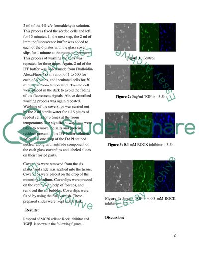
?Immunostaining and Microscopic Analysis of Adherent Cells Introduction: In the past Biologist were confined to the traditional transmission electron microscope and correlated the morphology with molecular and biochemical data. Use of the electron microscopy provides details in changes of cells due to ecological effects on living cells. Fluorescence emission in the fluorescence microscopy is not only characterized by the position and intensity but also the wavelength, lifetime and polarization. Use of fluorescence imaging has made it easier the observation of photo physical events. Rotation and mobility of the fluorescence is measured through the TR-FAIM as “Time-resolved fluorescence anisotropy imaging”. New applications are used to find the path of the unexpected discoveries (Suhling, French and Pillips 2004). According to Haupt, Pelling and Horton (2006) the detection of material subjected to damage the cells is a valuable strategy for both researchers and manufacturers of scientific equipments. A novel assay is required to study the Immunostaining and analysis of adherent cells by using the microscopic developments. This study reveals the analysis of adherent analysis by the treatments of inhibitors and other important fluorescence material (Vincent and Maiese 1999). Khosarvi-Fa et al (2008) determined the fixative solution used on the monolayer for the adherent cells DeMedeiore and Aho (2009) introduced the detection of intercellular agents on the surface of the adherent cells. Some other scholars also emphasized on the Immunostaining and analysis of the adherent cells. Use of the confocal microscopy has become common as non-ionizing radiations are employed, which are also used for the tissue preparation and study of the living cells (Varon et al., 2008). TGF? performs the dual role as metastasis promoter and tumour suppressor and keeps the balance between Smad3 and Smad2. Smad2 is found to be deleted or mutated in human cancers (Stuelten et al., 2007). Aim of this work lies to assay the adherent cells by using the process of Immunostaining and analysis through microscope. Materials and Methods: Precautionary steps were taken to protect the human body and avoiding the solution splashing. The MG-63 cells from the osteoblast were taken, and seeded these cells on the glass coverslips of 22mm x 22mm at a low density of 5000 and cells placed in each coverslips. The set up of the 6 plates was prepared for this experiment. These cells were treated with a 0.3 ROCK inhibitor M H1152 and five ng/ml TGF?. These treatments were made in the Rosewell Park in a normal growth medium of 10% v/v Foetal Calf Serum, and 1% Penicillin (Streptomycin). Each well of the prepared six plates having the seeded cells was added 2 ml of the 4% v/v formaldehyde solution. This process fixed the seeded cells and left for 15 minutes. In the next step, the 2 ml of immunofluorescence buffer was added to each of the 6 plates with the glass cover slips for 1 minute at the room temperature. This process of washing the cells was repeated for three times. Again, 2 ml of the IFF buffer was added made from Phalloidin-AlexaFluor-488 in ration of 1 to 500 for each of 6 plates, and incubated cells for 30 minutes at room temperature. Treated cell were placed in the dark to avoid the fading of the fluorescent signals. Above described washing process was again repeated. Washing of the coverslips was carried out by the 2 ml sterile water for all 6 plates of seeded cells for 3 times at the room temperature. The significant washings were taken to remove the salts and protein residues present in the IFF buffer solution. After this, one drop of the DAPI stained nuclear along with antifade component on the each glass coverslips and labeled slides on their frosted parts. Coverslips were removed from the six plates, and slide was applied into the tissue. Coverslips were placed on the drop of the mounting medium. Coverslips were pressed on the centre with help of forceps, and removed the air bubbles. Coverslips were fixed by using the nail varnish. These prepared slides were kept in the dark. Results: Respond of MG36 cells to Rock inhibitor and TGF? is shown in the following figures. Figure 1: Control Figure 2: 5ng/ml TGF-b – 3.5h Figure 3: 0.3 mM ROCK inhibitor – 3.5h Figure 4: 5ng/ml TGF-b + 0.3 mM ROCK inhibitor – 3.5h Discussion: The stimulation of the cultured cells was carried out with the transforming factors known as the TGF-? in the current work. This TGF-? was soluble and produced by the cells and stored in the same cells of the extracellular matrix. The TGF-? was bind to the cells of the surface receptor and multiple signalling paths were activated. Activation of signalling paths resulted into a directed and a wide range of the cellular functions. It has been seen that production of extracellular matrix (ECM), cells proliferation, and cells migration was controlled by the TGF-?. An increased in the Migration or motility of cells is an important function of the metastatic cancer. This condition is only possible when disease spreads to other tissues of the body parts. In the current work, ROCK inhibitor M H1152 was used as the inhibitor of the cellular enzyme. This is the key effecter of the TGF-? during the period of the promotion of the EMT. Activation of the ROCK inhibitor resulted into the regulatory of myosin light chain that acted as motor part and provided a contractile response for the “”actin cytosleleton” at the rear (back end) of the moving cells forwardly. A contractile response supported in propelling of the cells forward as through a disassembly produced from adhesion of cell-matrix. The disassembly mechanism for the cell adhesion and cell-matrix was also seen. Adhesion disassembly and cells contractility were promoted. This finding also confirmed the report of (Ezratty et al., 2005) as they identified the downstream target for the microtubule adhesion assembly in the fibroblasts. It was significant when determined the activation of Rock inhibitor during the tail retraction or de-adhesion of the rear cells. Confocal microscopy and immunoflourescent of the cells treated with the ROCK inhibitor showed dramatic accumulation of adherent cells in a large number of untreated tails. This confirmed that endosomes of cells were more accumulated in the motility process or in the migrating cells. It also showed that a higher trafficking of endosomes occurred during the cell migration. In the control culture as shown in figure 1 no changes were detected in the overall period of the experiment. This finding is also verified from the work of Bursch et al., (2000). This control demonstrated the typical cytokeratin patterns. Control cells were apparent as shown in the figure 1. There are three forms of TGF?s, which have similar properties and associate with the development of the fibrosis. In the current study, the TGF? was significantly located and suggested that cytokine had effects on its functions. These features were also confirmed by the research work of Higgins et al., (2009). From the figure 2, it is clear that cell treated with 5ng/ml TGF ? -b showed for 3.5 hours. For cells, 0.3 mM ROCK inhibitor was used for treatment of cells for 3.5 hours as shown in figure 3. The properties of cells were demonstrated as given in figure 3, when 0.3mM ROCK inhibitor was applied the cell nucleus were more apparent as compared to cells in figure 2 treated with 5ng/ml TGF-b for the same time of 3.5 hours in both cases. ROCK Inhibitor protected the nucleus from the damage. This finding was also revealed in the work of Yamashita et al., (2007). When cells were treated with 0.3mM ROCK inhibitor – 3.5h the results showed a clear image of the nucleus as shown in figure 4. The results revealed that ROCK inhibitor reduced the apoptosis. In this work, the cell nucleus was visualized with the same time of the actin cytoskeleton. By using the 4',6-diamidino-2-phenylindole, the cell nucleus was visualized that reflected the fluorescent stain. The wavelength of the 4', 6-diamidino-2-phenylindole absorption ranged between 358 nm (ultraviolet) and 461 nm (blue). Fluorescent Microscopy resulted into 4, 6-diamidino-2-phenylindole visualization through the blue/cyan filter. Earlier study also characterised the fluorescence based on the absorption and other fluorescent properties. The fluorescence was expressed as the ratio between the number of emitted photons and number of absorbed photons. The fluorescent Microscope visualised the actin cytoskeleton and properties of Phalloidin. This Phalloidin as a potent toxin that was taken from the death cap of the mushrooms. Chromatin condensation was also visualized by the use of the conformal microscopy. This is evident from the study of Tatton and Rideout (1999) who found that Parkinson disease resulted into death of the neuron cells via the apoptosis. Structural detail of nucleus was provided by the identification of the apoptotic nuclei through confocal microscopy. Cells treated with the apoptosis inducers showed the condensation of the chromatin material by using the fluorescence microscope. Nuclei of the cells also demonstrated the staining. Those cells, which had undergone apoptosis, loosed the nucleolar staining. Conclusions: The outlook of the fluorescence microscopy and its applications in the biomedical sciences is bright. Development of confocal microscopy demonstrated the greater precision in manipulation of cells and molecules. In conjunction with the demands of immunoflourescent dyes, the imagings of the living cells have shown more progress in bio-medical sciences. Study of the cell structure such as nucleus and other cell organelles is now easier with the help of Immunostaining technique by using the microscopic instruments. Visualisation of the Actin Cytoskeleton was also observed with several related results under the use of Phalloidin. Cellular integrity of live, dead as well as apoptosis is monitored through the fluorescent dyes. Effeects of the inhibitors and TGF? family is also most important as discussed in the current study. References: Bursch, W., Hochegger, K., Torok, L., Marian, B., Ellinger, A., and Hermann, R. (2000). “Autophagic and apoptotic types of programmed cell death exhibit different fates of cytoskeletal filaments”, Journal of Cell Science 113, P.P. 1189-1198 DeMedeiore, R. and Aho, J. (2009). “Immunostaining protocols for flow cytometric analysis of adherent cells”, The International Journal of Life Science Methods. Ezratty, E.J., M.A. Partridge, and G.G. Gundersen. 2005. Microtubule-induced focal adhesion disassembly is mediated by dynamin and focal adhesion kinase. Nat. Cell Biol. 7, p.p.581–590. Higgins, D., Basaraba, R., Hohnbaum, A., Lee, E., Grainger, D., Gonzalez, J. (2009). “Localized Immunosuppressive Environment in the Foreign Body Response to Implanted Biomaterials”, Am J Pathol. 175(1): p.p.161–170. Haupt, B., Pelling, A., and Horton, M. (2006). “Integrated Confocal and Scanning Probe Microscopy for Biomedical Research”, The Scientific World JOURNAL 6, P.P.1609–1618. Khosarvi-Far, R., Zakeri, Z., Lockshin, R., and Piacentini, M. (2008). Programmed Cell Death, Elsevier Inc. Stuelten, C., Kamarju, A., Wakefield, L., Roberts, A. (2007). “Lentiviral reporter constructs for fluorescence tracking of the temporospatial pattern of Smad3 signaling”, BioTechniques 43. p.p. 289-294. Suhling, K., French, P., and Phillips, D. (2004). Time-resolved fluorescence microscopy, The Royal Society of Chemistry and Owner Societies 2005. Tatton, N.A., and Rideout, H.J. (1999). “Confocal microscopy as a tool to examine DNA fragmentation, chromatin condensation and other apoptotic changes in Parkinson's disease.”, Parkinsonism & Related Disorders. Volume 5, Issue 4 , p.p.179-186 Varon, C., Rottiers, P., Enzan, J., Reuzeau, E., Basoni, C., Krame, Genot, E. (2008). “TGF?1 regulates endothelial cell spreading and hypertrophy through a Rac–p38-mediated pathway”, Biol. Cell. 100, p.p.537–550. Vincent, A., Maiese, K. (1999). “Direct Temporal Analysis of Apoptosis Induction in Living Adherent Neurons”, J Histochem Cytochem, vol. 47 no. 5 p.p.661-671 Yamashita, K., Kotani, Y., Nakajima, Y., Shimazawa, M., Yoshimura, S., Nakashima, S., Iwama, T., and Hara, H. (2007). “Fasudil, a Rho kinase (ROCK) inhibitor, protects against ischemic neuronal damage in vitro and in vivo by acting directly on neurons”, Elsevier, Vol. 1154, p.p. 215-224. Read More
Cite this document
  • APA
  • MLA
  • CHICAGO
(“Application of Microscopy in Biomedical Sciences Lab Report”, n.d.)
Application of Microscopy in Biomedical Sciences Lab Report. Retrieved from https://studentshare.org/health-sciences-medicine/1469148-application-of-microscopy-in-biomedical-sciences
(Application of Microscopy in Biomedical Sciences Lab Report)
Application of Microscopy in Biomedical Sciences Lab Report. https://studentshare.org/health-sciences-medicine/1469148-application-of-microscopy-in-biomedical-sciences.
“Application of Microscopy in Biomedical Sciences Lab Report”, n.d. https://studentshare.org/health-sciences-medicine/1469148-application-of-microscopy-in-biomedical-sciences.
  • Cited: 0 times

CHECK THESE SAMPLES OF Immunostaining and Microscopic Analysis of Adherent Cells

Electron Microscopy

'Prelims', Electron Microscopy and analysis, 1:1, 0 – 19.... 'Prelims', Electron Microscopy and analysis, 1:1, 0 – 19.... This essay discusses electron microscopy, that is a technique in microscopy that offers higher magnification and greater depth of field in viewing objects....
4 Pages (1000 words) Coursework

Atomic Force Microscope

Like the first group of authors, the authors of this article, also employed experimental design and the quantitative method techniques in the analysis of their research.... Name Institution Date Article Review The article by Anshu Bagga, Mathur, Geoge, Monty Reichert, and Tuskey deals with the study of force transmission in endothelial cells using the Atomic Force and the Total Internal Reflection Fluorescence microscopy.... In the second article, the authors Mike, Harton and Guillaume, are more concern with analyzing the atomic force microscope in ascertaining magnitude and the nature of strains upon which cells often respond....
3 Pages (750 words) Coursework

Transforming Growth Factor-beta and ROCK Inhibitor in Immunostaining

Lab Report: Transforming Growth Factor-beta and ROCK Inhibitor in Immunostaining and Microscopic Analysis of Adherent Cells Name Institution.... Immunostaining and Microscopic Analysis of Adherent Cells deals with substrates that are inexpensive and have genetics that are well characterized, and has an increasingly increased number of mutant host strains and cloning vectors.... This report examined the transforming growth factor-beta and ROCK inhibitor in Immunostaining and Microscopic Analysis of Adherent Cells....
7 Pages (1750 words) Lab Report

Application of Microscopy in Biomedical Sciences

Use of the electron microscopy provides details in changes of cells due to ecological effects on living cells.... ome other scholars also emphasized the importance of confocal microscopy used for the observation of living cells.... Use of the confocal microscopy is now common as non-ionizing radiations are employed, which are also used for the tissue preparation and study of the living cells.... For the epithelial cells, the most important factor of family TGFβ is the TGFβ 1....
6 Pages (1500 words) Lab Report

Structure of Eukaryotic Cells

An essay "Structure of Eukaryotic cells" discusses the structure of eukaryotic cells and the importance of the membranes.... The eukaryotic cells have a great similarity to the prokaryotic cells which contain structures such as ribosomes, plasma membrane, and cytoplasm.... Eukaryotic cells are the most structurally advanced of the major cell types existing in form of complex cells in which genetic material is organized into membrane-bound nuclei....
5 Pages (1250 words) Essay

Cell biology

In this branch, the structure of cells, life.... , properties, interactions with the environment, function, division, death and organelles (their locations within cells, functions, structures, compositions and adaptations) found within cells are delved into.... The study of cells is done in a molecular level or is conducted suing Insert Cell biology, alternatively referred to as cytology, is an area of biology that deals with (studies) cells (Lodish, 348)....
1 Pages (250 words) Research Paper

Using a Compound Light Microscope

Through the use of microscopes, it has become easy to discover and learn about the microorganisms, structure of cells, and know more about animals, fungi, and plants more closely.... Moreover, light microscopes are used widely to look more closely at cells in tissue; however, its visibility is limited to see objects less than 0.... lectron microscopes are used to see much smaller objects which are inside cells.... This report "Using a Compound Light Microscope" discusses the study of the very small organisms and structures that require microscopic examination....
7 Pages (1750 words) Report

Techniques and Results Achieved from Scanning Electronic Microscopy and Transmission Electronic Microscopy

This paper seeks to analyze the different images observed and techniques during visualization of human red blood cells by use of TEM and SEM.... Air drying mainly results to the collapsing and shrinking; hence, drying is mainly achieved through replacing the water cells with organic solvents such as ethanol or acetone....
11 Pages (2750 words) Coursework
sponsored ads
We use cookies to create the best experience for you. Keep on browsing if you are OK with that, or find out how to manage cookies.
Contact Us